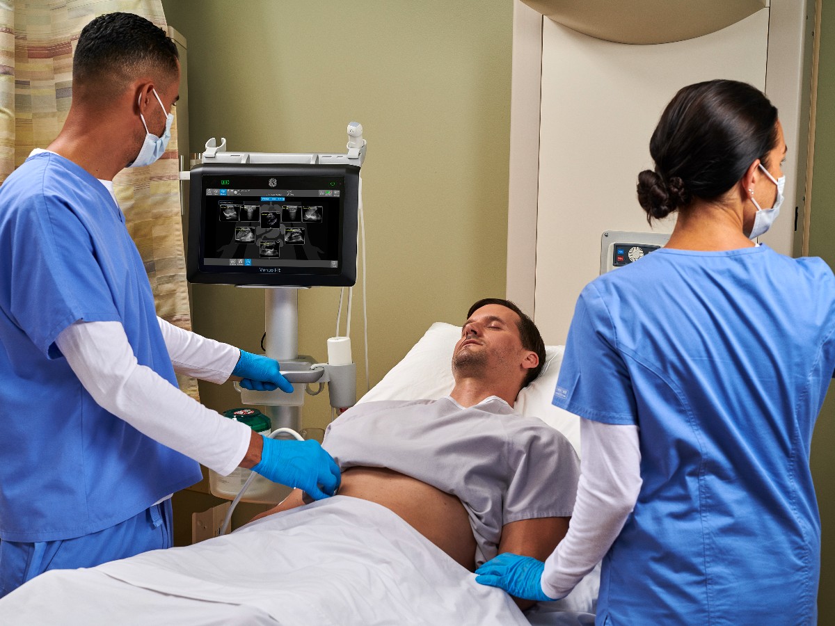
The use of point of care ultrasound and radiology ultrasound could be thought of as competing medical specialties. However, both of these imaging techniques have found their own place in medicine.
Ultrasound has a long history, but equipment restraints meant that it was only performed in hospitals' radiology departments until about 30 years ago. Once ultrasound machines became smaller, more portable, and cheaper, the use of POCUS took off, especially in the emergency department (ED) and critical care units.1 The introduction of POCUS has expanded the role of ultrasound at the bedside, providing numerous advantages for physicians, patients, and hospital systems.
Point of Care Ultrasound and Radiology: Workflow
To understand the advantages of POCUS, consider the workflows for both point of care ultrasound and radiology. POCUS is a bedside exam performed by the immediate supervising physician and read as the scan is being performed by that same physician. POCUS can be performed almost immediately.
Instead, radiology ultrasound requires the following sequence of steps:
-
The order is placed for a radiology ultrasound exam.
-
Typically, there is a wait time for radiology.
-
The patient is then transported to radiology, where a sonographer performs a scan.
-
The patient is then transported back to the original hospital location.
-
The radiologist reads the ultrasound scan and puts the report into the electronic medical record system.
Although this series of steps seems straightforward, this list doesn't factor in how busy radiology departments tend to be. Radiology exams are in high demand, leading to long wait times. By utilizing non-radiology physicians to perform POCUS exams, radiologists are free to perform the exams that require their expertise.3
The POCUS workflow introduces several efficiencies:
-
POCUS speeds up the scan and time to assessment.
-
The ultrasound is performed at the bedside. The patient avoids transport, and the bedside physician can incorporate POCUS findings immediately into their history and physical exam (HPI) findings—quality, severity, associated signs and symptoms, and duration. In turn, they can integrate these findings into their clinical decision-making.
-
It reduces the need to involve a second physician (the radiologist) in the patient's care, giving the radiology department more time to perform other specialized tests and exams.3
POCUS and radiology ultrasound may be similar in technique, but they are used very differently. POCUS helps answer a specific clinical question, such as "Does this patient with abdominal pain have gallstones?" It is a limited exam with a focus on answering just one clinical concern. Based on the results, the clinician can initiate different clinical pathways, such as admission and surgery consult for gallstones or signs of acute cholecystits versus further imaging if gallstones are not present.
Radiology ultrasound usually involves a more comprehensive exam, expanding the number and kind of clinical questions that physicians can answer for right upper quadrant (RUQ) pain. For example, in addition to scanning for gallstones, an abdominal ultrasound exam can evaluate for liver and pancreatic lesions if gallstones are absent, as these can both also cause RUQ pain.
To further explore point of care ultrasound and the radiology ultrasound exam, consider a hypothetical clinical scenario.
Case Study: Point of Care Ultrasound and Radiology
It is 7 p.m. on a Saturday, and a 36 year-old woman comes to the ED complaining of abdominal pain. On HPI, the patient states that she has been having right-sided abdominal pain for a few days that has become persistent and unrelenting over the past few hours. The patient also complains of nausea and lack of appetite. Upon exam, it is determined that she has pain in her RUQ. The differential diagnosis for this patient is broad, but the ER physician suspects gallbladder disease based on HPI and physical exam and puts in an order for lab tests, an ultrasound, and a CT scan.-
The lab results come back showing leukocytosis, bilirubin of 2 mg/dL, and normal lipase levels. After the lab results return, the physician has an even stronger suspicion that the patient has acute cholecystitis.
Since radiology is busy, it will be hours before the patient can get an ultrasound or CT scan. This is not unusual in many hospitals. One of the most common reasons for slow ED turnaround time is waiting for imaging—an average turnaround time for a CT scan is 5.9 hours.5
The physician instead decides to perform a POCUS exam of the patient's abdomen. If it indicates acute cholecystitis, the physician can consult surgery immediately. If the scan is negative, the doctor can schedule a CT scan.
Within 10 minutes, the ED doctor in our example scans the patient's abdomen at the bedside2, and finds gallstones and gallbladder thickening, thereby confirming the clinical suspicion that leads to a surgery consult. Additionally, the use of POCUS has allowed the patient to avoid a CT scan and exposure to radiation.
Detecting Gallbladder Disease
Twenty million patients are affected by gallbladder disease in the US every year. Unfortunately, gallbladder disease can't be accurately diagnosed based on HPI alone, necessitating that patients undergo further imaging for a definitive diagnosis. Ultrasound can often provide helpful information in evaluating quickly for gallbladder disease.4
Traditionally, biliary ultrasound has required a trip to radiology. However, recent studies have shown that POCUS in the ED has the potential to accelerate diagnosis and treatment in patients with gallbladder disease.2 In fact, a meta-analysis showed that POCUS surpassed all other bedside modalities in sensitivity while diagnosing of biliary disease and that it is similarly accurate to the findings from radiology-diagnosed biliary disease.2
Despite the fact that biliary POCUS is both as sensitive and specific as radiology ultrasound, many surgeons want confirmatory biliary radiology ultrasound, even if POCUS shows cholelithiasis or acute cholecystitis.2 Hilsden, et al., has confirmed that the presence of gallstones found on a POCUS scan in a patient with a significant suspicion for biliary pathology is highly predictive of the eventual need for a cholecystectomy.2
Since time to diagnosis and discharge is intimately associated with ED wait times, POCUS can introduce broader efficiencies while imaging suspected biliary disease. There is a significant difference in the length of the ED stay between patients who undergo biliary POCUS and those who are referred for radiology ultrasound. Patients who underwent radiology ultrasound were in the ED for an average of 433 minutes (± 50 minutes). In contrast, POCUS patients had an average discharge time of 309 minutes (±30 minutes) after admission.2 This difference may help alleviate delays in care and ED crowding.
The POCUS exam and radiology ultrasound exam are undeniably similar in performance. However, rather than create turf wars over who carries out the exams, POCUS can help streamline clinical processes for everyone. By assisting in diagnosis and decreasing wait times, POCUS frees up radiologists to perform exams on cases that may be more clinically complex and require a more comprehensive exam.
References
- Phillips L, Hiew M. Point of care ultrasound: Breaking the sound barrier in the emergency department. Australas J Ultrasound Med. 2019 Feb 19;22(1):3-5. doi: 10.1002/ajum.12129. PMID: 34760529; PMCID: PMC8411775. https://www.ncbi.nlm.nih.gov/pmc/articles/PMC8411775/
- Hilsden R, Leeper R, Koichopolos J et al. Point of care biliary ultrasound in the emergency department (BUSED): implications for surgical referral and emergency department wait times. Trauma Surgery and Acute Care Open. July 2018; 3(1) https://tsaco.bmj.com/content/3/1/e000164
- Hashim A, Junaid Tahir M, Ullah I. The utility of point of care ultrasound. Annals of Medicine and Surgery. November 2021. https://www.sciencedirect.com/science/article/pii/S2049080121009328?via%3Dihub
- Gallaher J, Charles A. Acute cholecystitis: a review. Journal of the American Medical Association. March 2022; 327(10):965-975 https://jamanetwork.com/journals/jama/fullarticle/2789654
- Perotte R, Lewin G Tambe U, et al. Improving emergency department flow: reducing turnaround time for CT scans. AMIA Annual Symposium Proceedings. December 2018; 897-906. https://www.ncbi.nlm.nih.gov/pmc/articles/PMC6371246/

