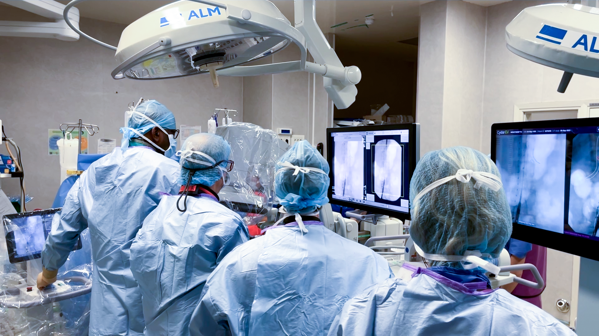
OEC Elite CFD paired with Cydar Maps image fusion system
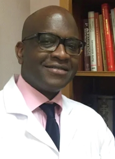
Interview with Pr. Elixène Jean-Baptiste, vascular surgeon, Head of the department of surgery and vascular medicine, regional reference center of ‘SOS aorta’ project for PACA-Est region, at the Nice University Hospital, France.
The department of surgery and vascular medicine of Nice University Hospital deals with all arterial diseases, venous peripheral vascular diseases as well as vascular access for hemodialysis. The Nice University Hospital has a complete endovascular technical platform, a high-performance vascular functional exploration laboratory and a non-stop technical care service.
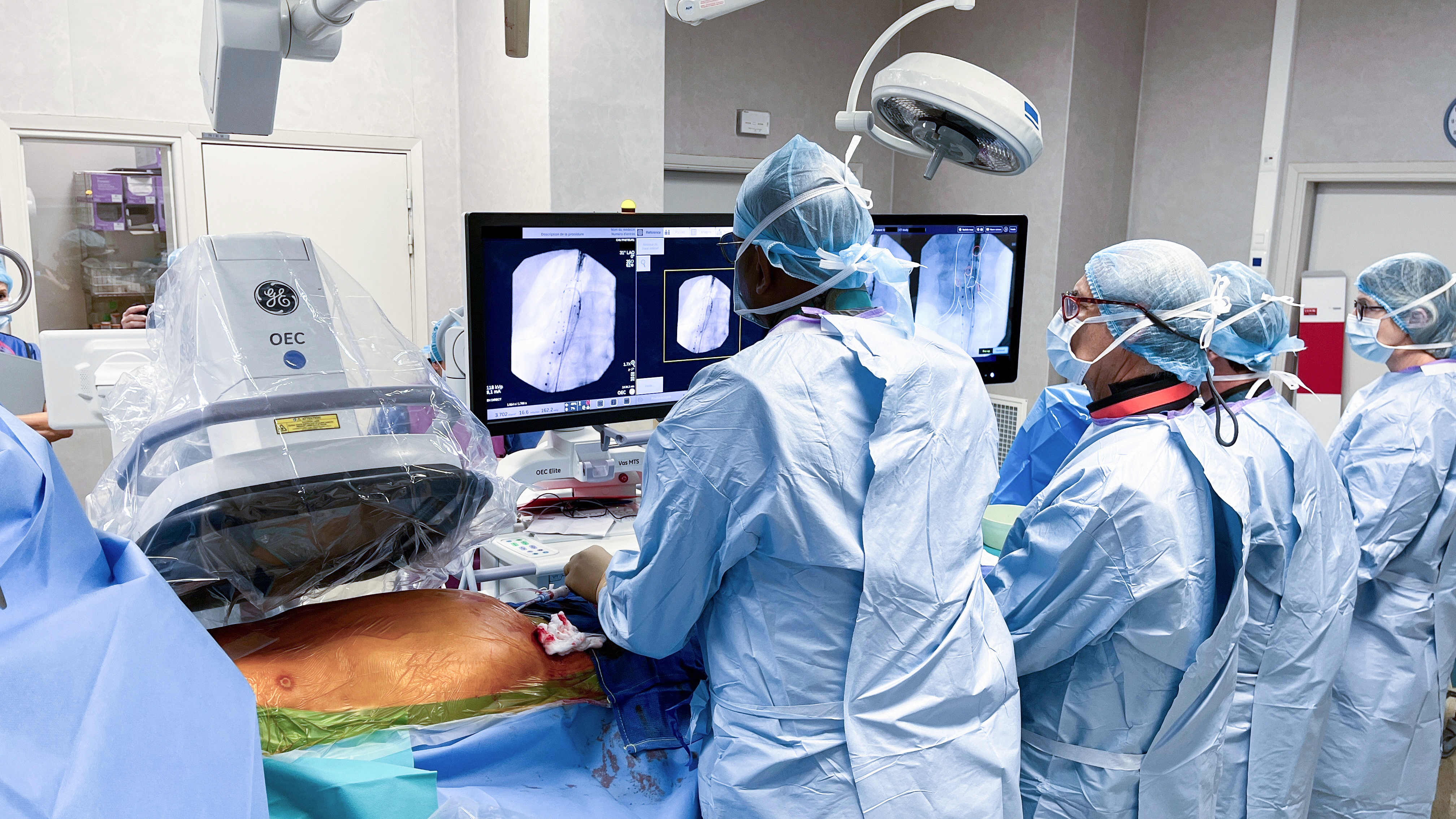
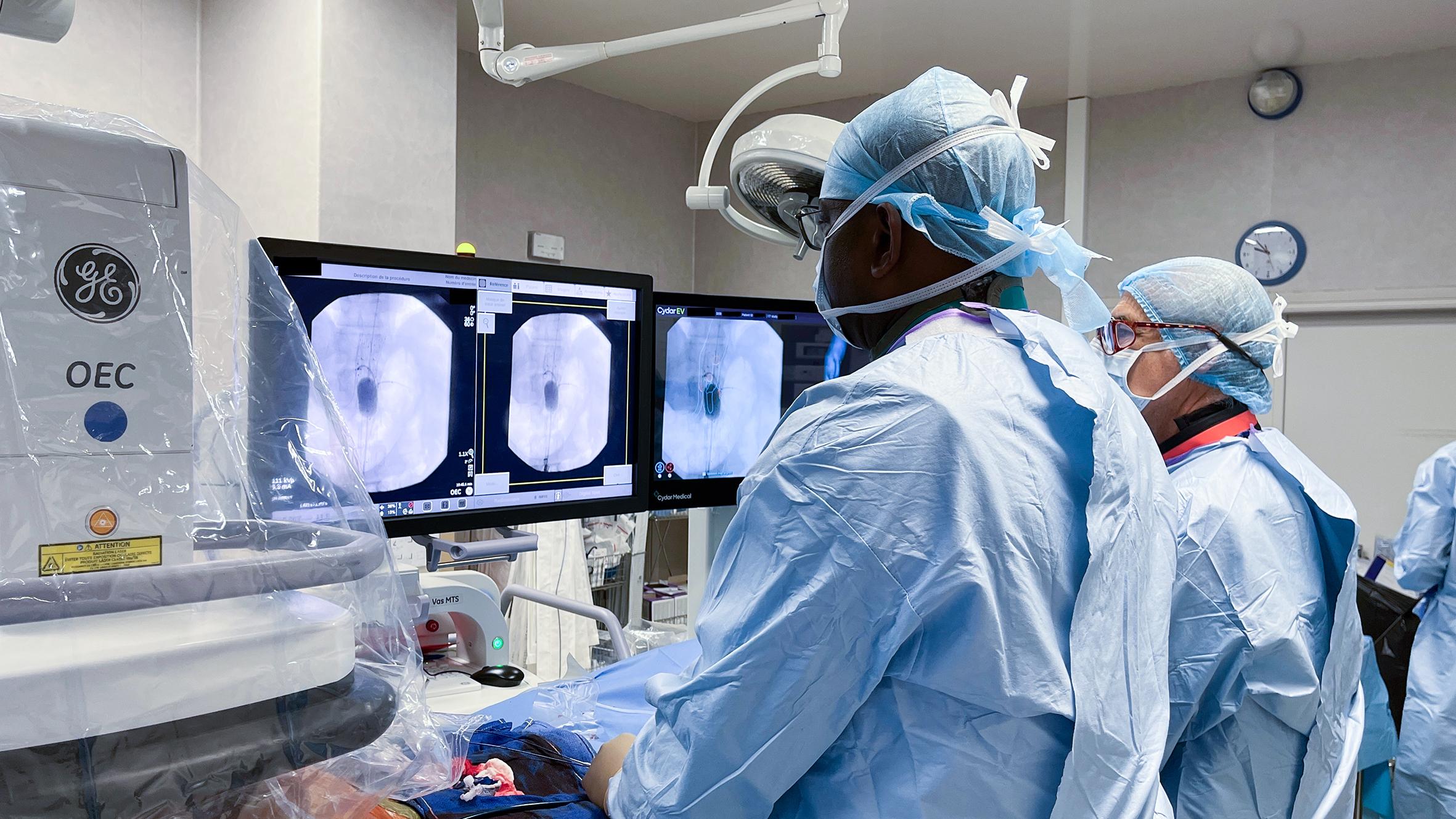
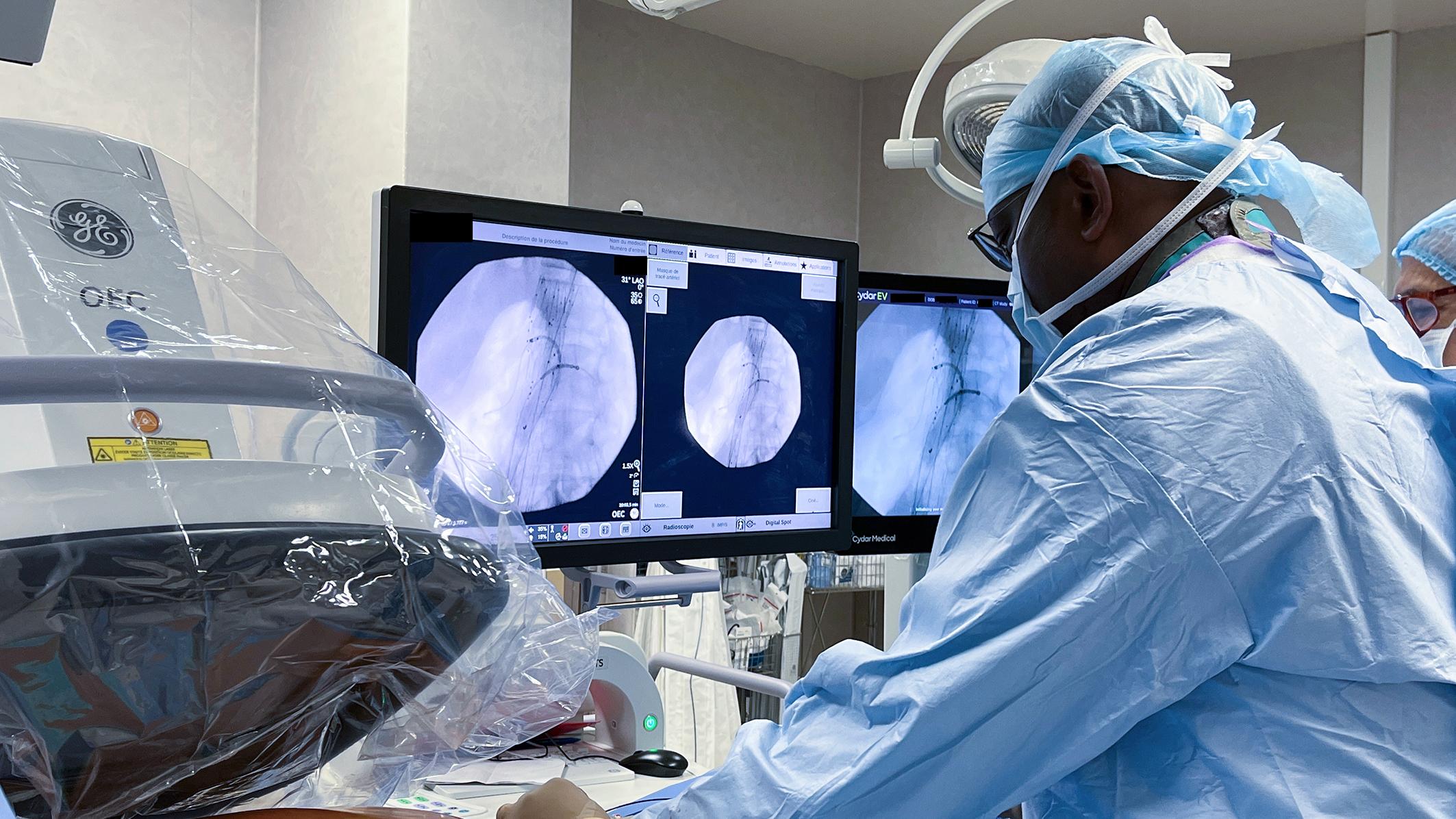
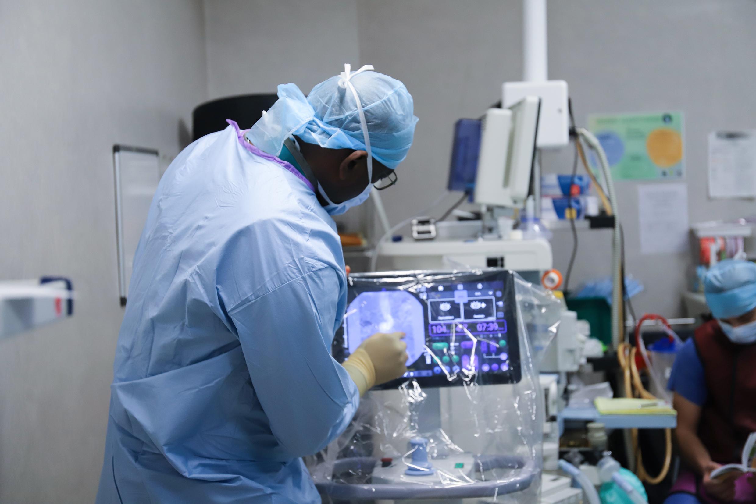
As the regional reference center for ‘SOS aorta’ project for PACA-EST region, that is creating a coordinated patient care pathway taking in charge acute aortic emergencies, Nice University Hospital has equipped a dedicated operating room (OR) with Cydar Maps (CYDAR Medical, Cambridge, United Kingdom) software. This cloud-based technology overlays the vascular mask from a preoperative three-dimensional computed tomography angiography (CTA) over intraoperative live fluoroscopy.
To be paired with this high-tech solution, the OEC Elite CFD motorized drive MD C-arm has been selected to provide imaging guidance of endovascular tools during endovascular aortic repair (EVAR) procedures.
Professor Jean-Baptiste, please tell us why you chose vascular surgery and what is your background?
"Since young, I have always wanted to become a surgeon. After my training in general surgery, I chose a fellowship in cardiovascular surgery. Then, vascular surgery attracted me the most, especially in relation to its possibilities for innovation in endovascular techniques. Vascular surgery is a specialty that can be labeled as ’young’ even if it is currently reaching a phase of maturity. There are enormous opportunities for innovations in endovascular surgical techniques with many new tools for these techniques. Regrettably, this doesn’t necessarily apply to some other surgical specialties which have already matured, ceased evolving, and are now losing prominence to the benefit of certain other medical specialties.
In France, we are fortunate that the vascular surgery community gives practitioners the opportunity to practice both endovascular surgical techniques as well as conventional surgical techniques also called ‘open’ surgery techniques.
So, after 5 years of training spent in general surgery, I pursued 2 years of specialization in vascular surgery. Then I completed a 3-year dual fellowship in vascular surgery at Nice University Hospital and at Lille University Hospital. At that time in Lille, we were doing endovascular procedures in an operating room equipped with one of the first vascular motorized mobile C-arms, OEC Elite 9900. This was before the era of hybrid rooms. After completing my Ph. D in Lille, I returned to Nice University Hospital working as a practitioner in vascular surgery. I am the head of the department of surgery and vascular medicine since 2015."
What is your volume of procedures and what benefits does OEC Elite CFD bring to them?
"The department of vascular surgery deals with all arterial disease cases, venous peripheral vascular disease cases as well as cases of vascular access for hemodialysis. We perform around 2000 procedures per year and out of this volume of procedures, about 60 to 80% are executed by endovascular approach. Concerning aortic procedures specifically, we perform about 60 to 80 interventions per year of which 60% with endovascular approach, and 40% with conventional surgery.
Fluoroscopic imaging is an essential tool during endovascular surgical procedures. In the future, for some procedures we may use alternative tools such as image-guided echography, but so far, fluoroscopic imaging has allowed the development of endovascular techniques and the growth of the vascular surgery specialty. Both endovascular and fluoroscopic imaging techniques are continuously being developed together in parallel.
“When we started to look for a new C-arm for our department, I asked for three important things for my practice: a motorized C-arm, the ability to pilot the C-arm by myself from commands on the table side or a satellite control station, and good image quality during endovascular procedures.”
Pr. Elixène Jean-Baptiste
When we started to look for a new C-arm for our department, I asked for three important things for my practice: a motorized C-arm, the ability to pilot the C-arm by myself from commands on the table side or a satellite control station, and good image quality during endovascular procedures.
Hence, we opted for the OEC Elite CFD motorized C-arm with the vascular set-up software “Vascular MTS”. When operating the commands of the OEC Elite CFD C-arm, we alternate between the Remote User Interface (RUI) or the Touch Table Side cart depending on the procedure. For peripheral procedures (i.e. lower limbs recanalization procedures) I use the RUI for its compactness and its quick access to the main features.
“For peripheral procedures (i.e. lower limbs recanalization procedures) I use the RUI for its compactness and its quick access to the main features.”
Pr. Elixène Jean-Baptiste
For EVAR we set up OEC Elite CFD in Vascular imaging profile, 8 pulses per second and in low dose mode. I use the Live Zoom to increase the size of the image without changing the dose of radiation. As the majority of our patients with these pathologies have renal failure as well, we dilute iodine contrast media with a saline solution to a concentration of 50% to avoid iodine shock. The good image quality of the angiography enables us to work in roadmap mode or with the Cydar Maps image fusion software.
“For endovascular aortic repair (EVAR) procedures, I often use the advanced features such as Live Zoom and the Digital Pen, easy to activate from the Touch Table Side.”
Pr. Elixène Jean-Baptiste
As we often change the angle of the C-arm to obtain different images as we catheterize the collateral arteries of the aorta, we need to come back to these positions, and thus we often use the preset position functions to record
Right or Left Anterior Orbital (RAO, LAO) angles as well as the cephalad caudal
angles.
I can maneuver the C-arm angle from the Touch Table Side. This is a key advantage of having the OEC Elite CFD in our department because we have two operating rooms and only one radiologic technologist. On many occasions, I am comfortable to let our technician help in another operating room and operate the OEC Elite CFD C-Arm by myself.
What I truly appreciate about OEC Elite CFD, is it allows you to be autonomous!"
Can you explain to us the reason for pairing OEC Elite CFD with Cydar Maps?
"Image fusion tool is a step further in the evolution of endovascular techniques. This tool aims to reduce the number of X-ray images and the volume of iodine injected in the patient during EVAR procedures.
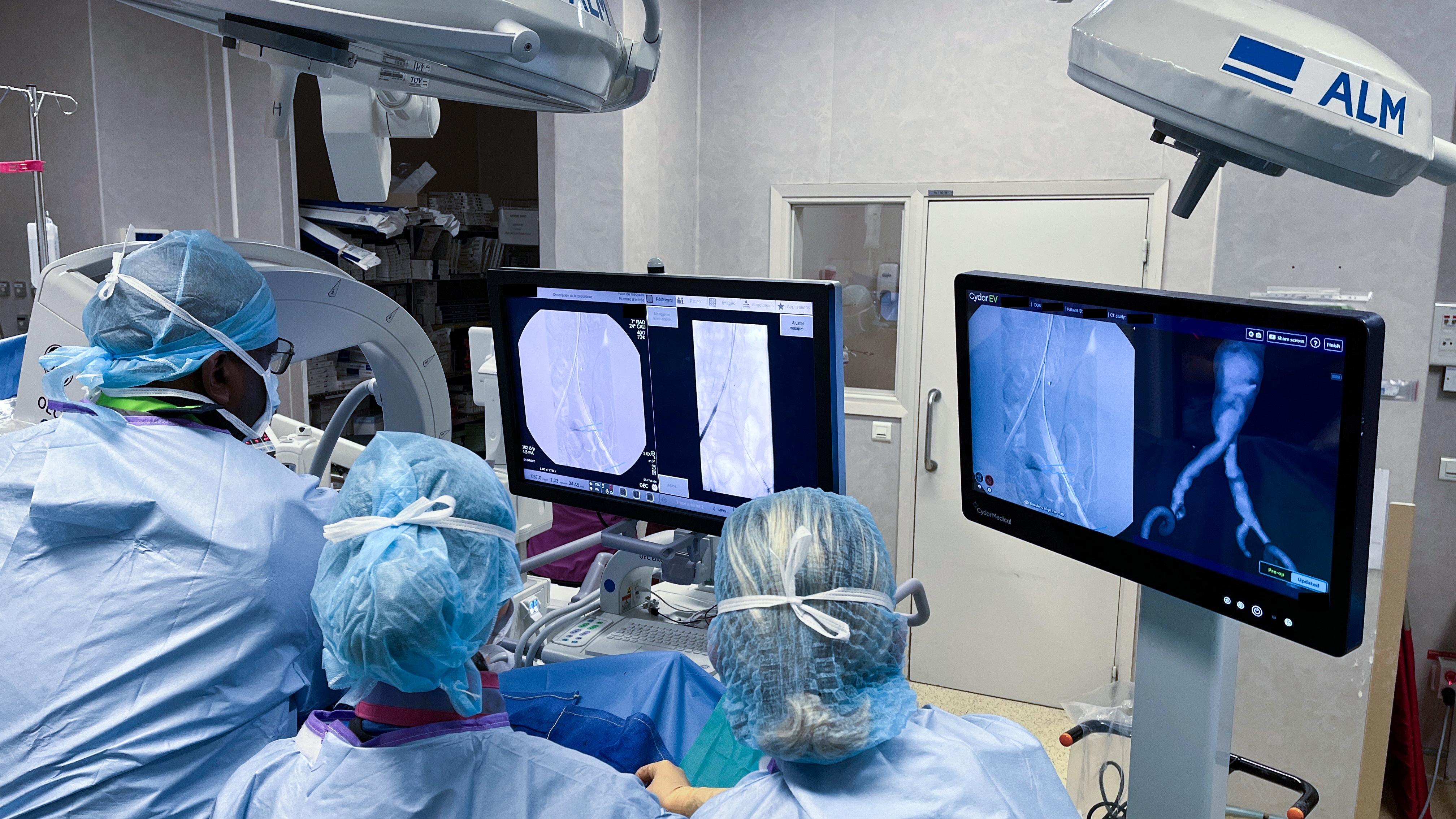
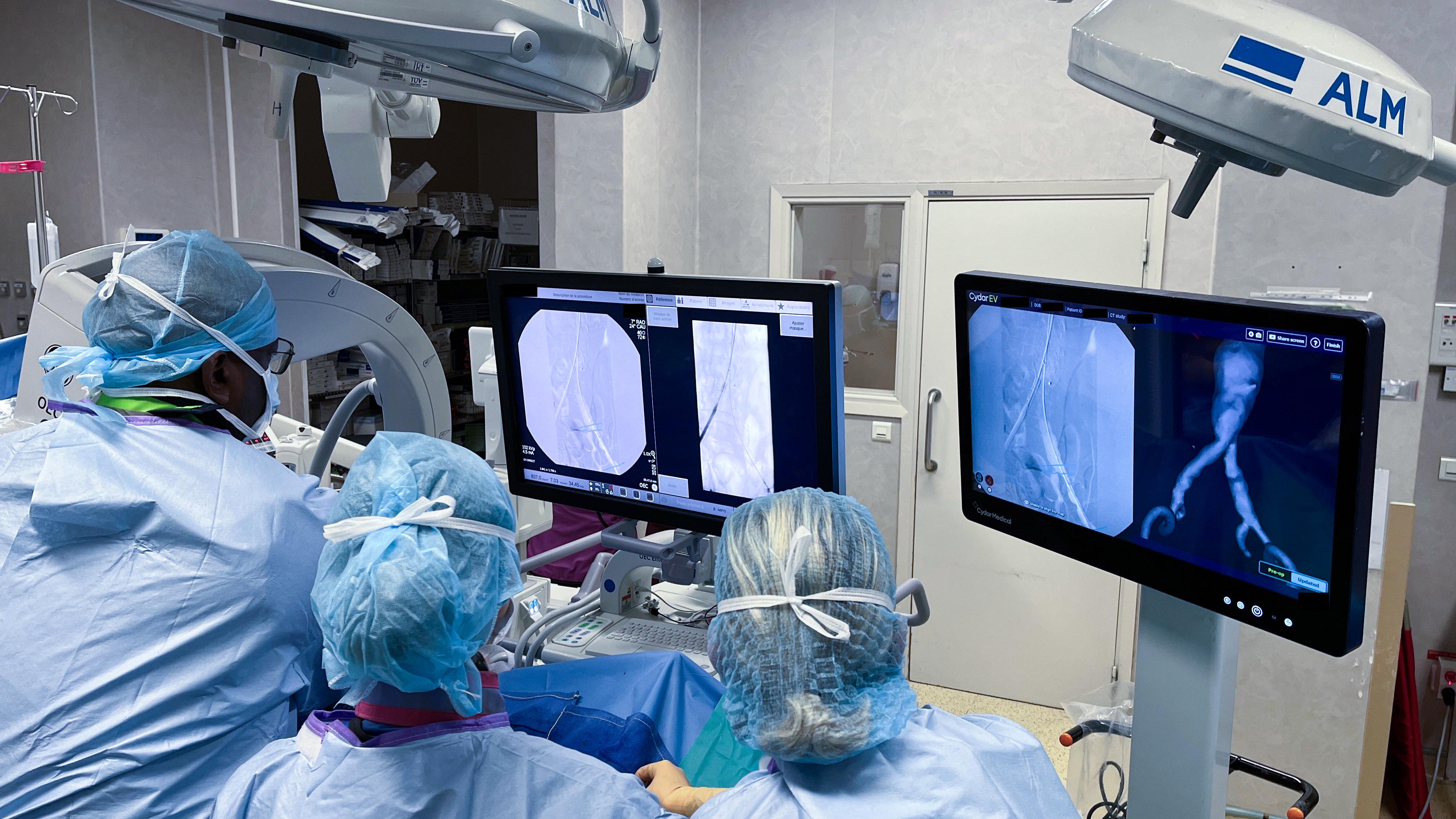
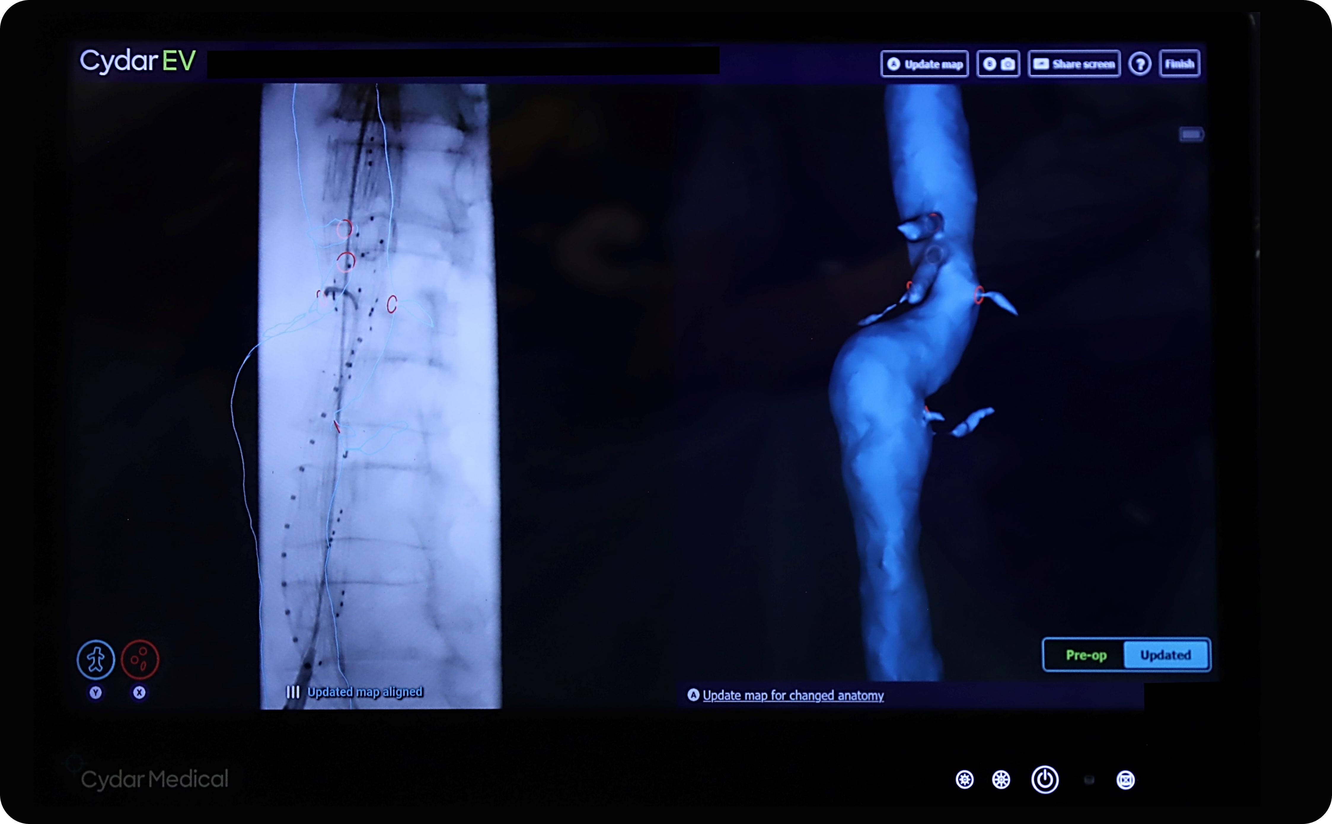
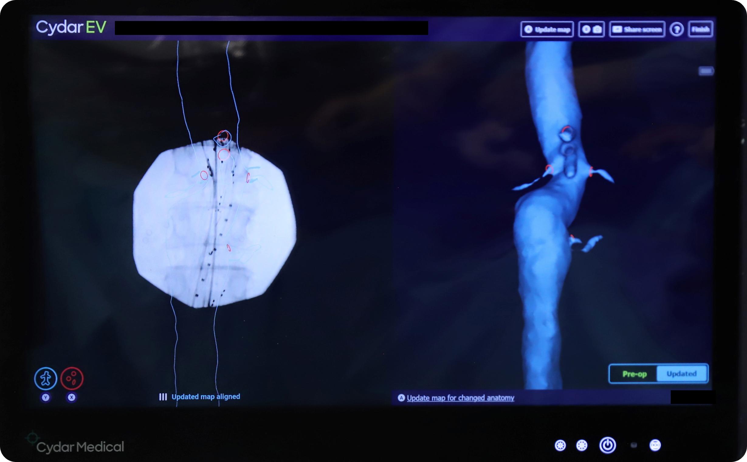
In simple words, Cydar Maps provides intraoperative imaging guidance by overlaying a preoperative vessel anatomy (or a 3D vascular mask) from the CTA of the patient on to intraoperative live fluoroscopy images. During the procedure, an artificial intelligence (AI) automatically corrects shifts between the 3D vascular mask and the patient’s anatomy, when we change the position of the patient table or the angle of the OEC Elite CFD C-arm.
Figure 1: Cydar monitor showing on the left the overlay of the CTA 3D vascular mask onto OEC Elite CFD live image
Figure 2: Cydar monitor showing on the left the overlay of the CTA 3D vascular mask onto OEC Elite CFD live image.
During the preparatory work before surgery, while working on the preoperative planning, I need to analyze the preoperative CTA scan. Cydar Maps helps me better reflect on the surgical strategy. I always explain to my interns that it is important to always have a flight plan before starting any endovascular procedure especially for complex procedures. The preparatory steps of the CTA image segmentation of the aorta and the step choice of the positioning of the rings on the mask, (a graphical representation of the ostia of collateral arteries) implement the surgical strategy into my thoughts. While defining the position of the ostium to be catheterized, I also decide the angle of the C-arm that matches the field of view I need for the catheterization of the considered artery. These choices are going to be important while executing the surgical gesture itself.
“What I truly appreciate about OEC Elite CFD, is it allows you to be autonomous! ”
Pr. Elixène Jean-Baptiste
During the surgery, the guidance by the overlay of the 3D vascular mask over the live fluoroscopy, reduces the chances to struggle to find the right fluoroscopic view for the catheterization of an arterial bifurcation. In addition, Cydar Maps automatically repositions the 3D mask over the live fluoroscopy in synchronization of eventual changes of anatomy coverage of the fluoroscopy. No action from the surgeon is required. As we don’t have engineers in the OR to support us, I really appreciate the additional autonomy I have during my surgery.
After the surgery, I can replay and quietly review with Cydar Maps the images of fluoroscopy with the 3D vascular mask of the different steps of a complex procedure and subsequently review how to overcome the potential difficulties encountered. It is a kind of rehearsal training tool for the surgeon."
In a nutshell, how do you qualify the clinical value of pairing OEC Elite CFD with Cydar Maps?
"We have been using OEC Elite CFD paired with Cydar Maps for more than a year now, during all our aortic repair procedures, simple and complex. This solution of combined advanced imaging technologies supports us to manage the radiation dose and the volume of iodine injected in the patient.
The combination of Cydar Maps preoperative planning software and the preset positions of OEC Elite CFD enables us to save time in the execution of the procedure: we no longer spend time finding the correct angulation to catheterize the ostium. The procedure is simplified, and we are less exposed to X-ray radiation.
The image quality of the OEC Elite CFD paired with the Cydar Maps’s 3D vascular mask with automatic repositioning reduces the number of iodine injections for patients with severe renal failure. We only need to perform one angiography at the beginning of the procedure to overlay the 3D vascular mask over the fluoroscopic image, and one angiography at the end of the procedure for the final control of the stent graft placement.
This technological solution brings autonomy, support, and confidence to the surgeon during his simple and complex endovascular procedures."
“The combination of Cydar Maps preoperative planning software and the preset positions of OEC Elite CFD enables us to save time in the execution of the procedure.”
Pr. Elixène Jean-Baptiste
|
|
Pr. Elixène Jean-Baptiste is a graduate vascular surgeon of Nice University of Medicine. He defended his Ph. D thesis in biomaterial research in 2012 after a dual fellowship at Nice University Hospital and Lille University Hospital. Currently based in Nice, he is at the head of the department of surgery and vascular medicine and the regional referent of ‘SOS aorta’ project for PACA-Est region, at the Nice University Hospital, France. |
The statements by GE’s customers described here are based on their own opinions and on results that were achieved in the customer’s unique setting. Since there is no “typical” hospital and many variables exist, i.e. hospital size, case mix, etc.. there can be no guarantee that other customers will achieve the same results. JB04064UK

