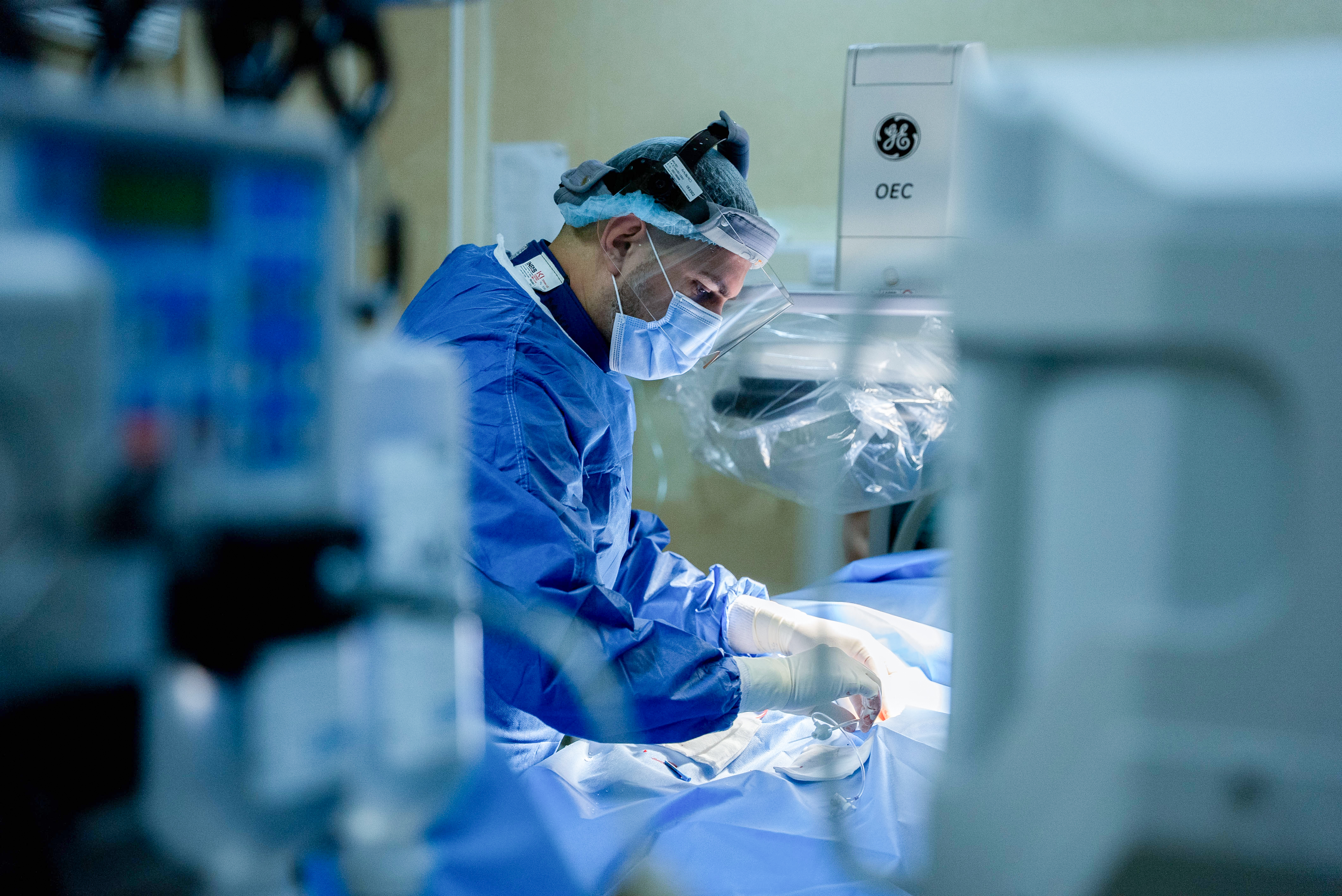
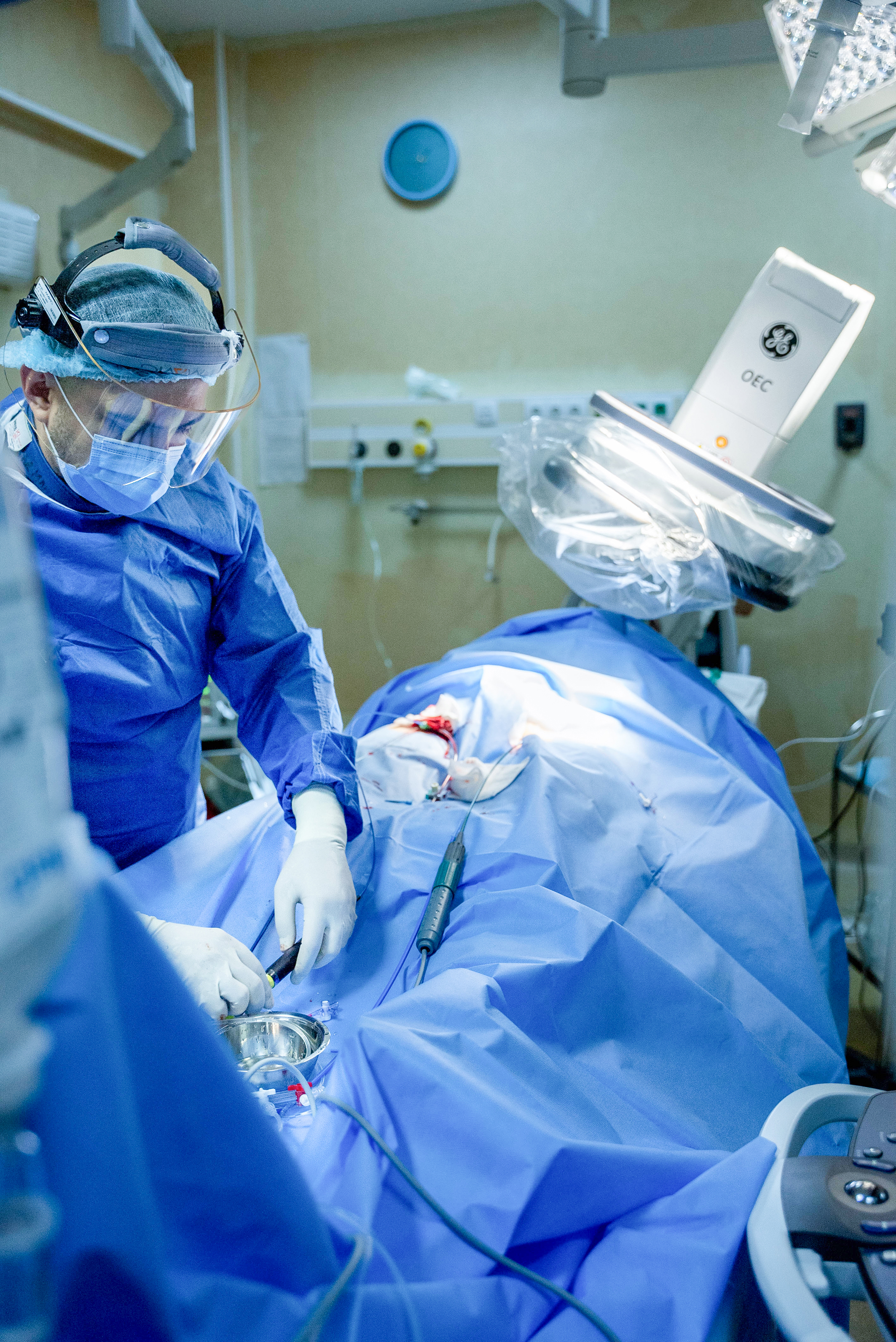
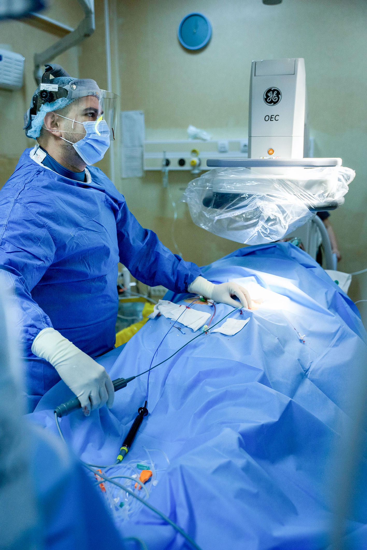
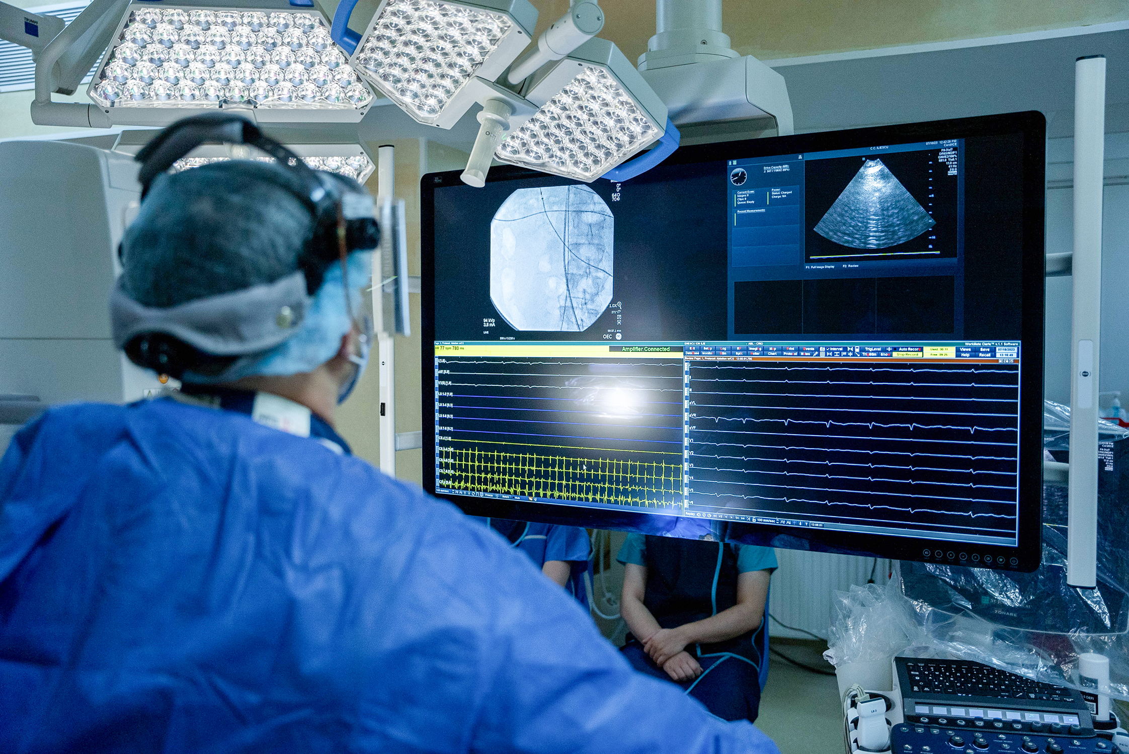
Interview with Dr. Sergiu-Nicolae Şipoş, Cardiologist and Interventional Rhythmologist, EP and Pacing Department, Institute for Cardiovascular Diseases, hospital CC Iliescu, Bucharest, Romania.
Can you describe CC Iliescu National Institute for Cardiovascular Diseases and your ongoing projects?
"We operate as a public university hospital in Romania, recognized as a tertiary center, signifying our commitment to delivering the highest level of healthcare. As a result, we fall under the administration of the Ministry of Health. Our facility specializes in performing procedures that may be less accessible in other centers, such as aortic artery dissection, complex coronary percutaneous dilatations and certain types of electrophysiology (EP) procedures, and device implantations. Patients from across the country, particularly from the southern region of Romania, seek our services.
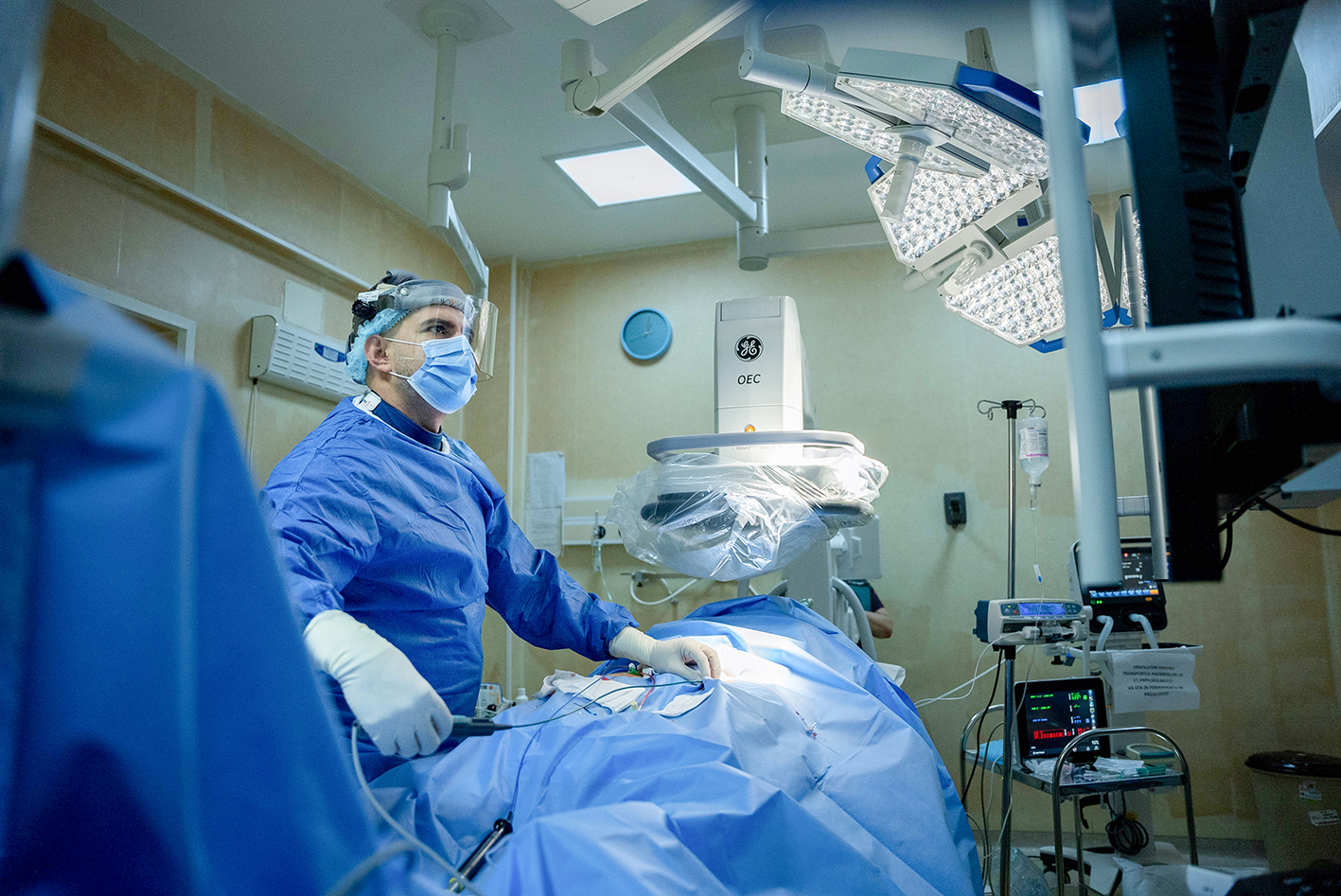
Our team consists of interventional cardiologists responsible for procedures like coronary angiography and stenting. Additionally, we have interventional rhythmologists who specialize in working with implantable devices, including pacemakers, as well as conducting electrophysiology and ablation procedures. As cardiologist with expertise in interventional rhythmology, I am part of the EP and Pacing Department, which comprises four physicians dedicated to EP procedures, pacing, devices and electrophysiology. As a high-volume center for Romania, conducting approximately 1,200 procedures annually.
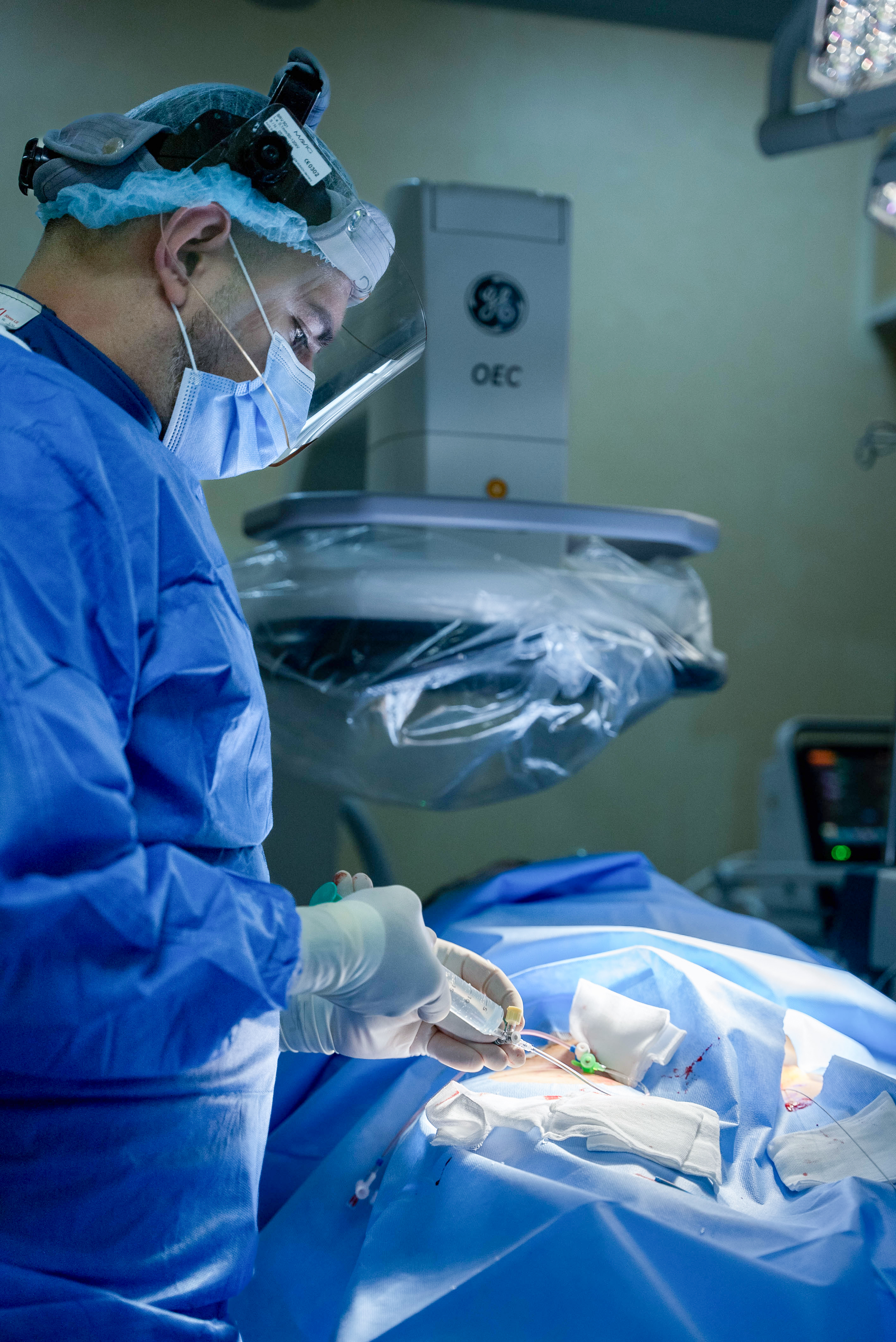
Regarding eletrophysiology (EP) procedures, we boast the highest procedural volume among all centers. We have two operating rooms. One is designed for implantable devices while the other is primarily used for EP procedures and complex devices. In that second room we have recently acquired an OEC Elite CFD mobile C-arm."
What types of procedures are you performing?
"The procedures we conduct are categorized as either ”simple” or ”complex” in accordance with the Ministry of Health’s criteria, which considers factors such as the volume of materials used and the complexity of the infrastructure required for the procedure.
For example, with simple devices like dual-chamber or single-chamber pacemakers, we rely solely on fluoroscopy.
However, for specific types of ablations in atrial fibrillation, ventricular tachycardias, atrial flutter for example, a 3D mapping system or a cryoablation console can also be necessary.
About half of the procedures we do are complex, and half are considered simple."
In what ways OEC Elite CFD does assist your procedures?
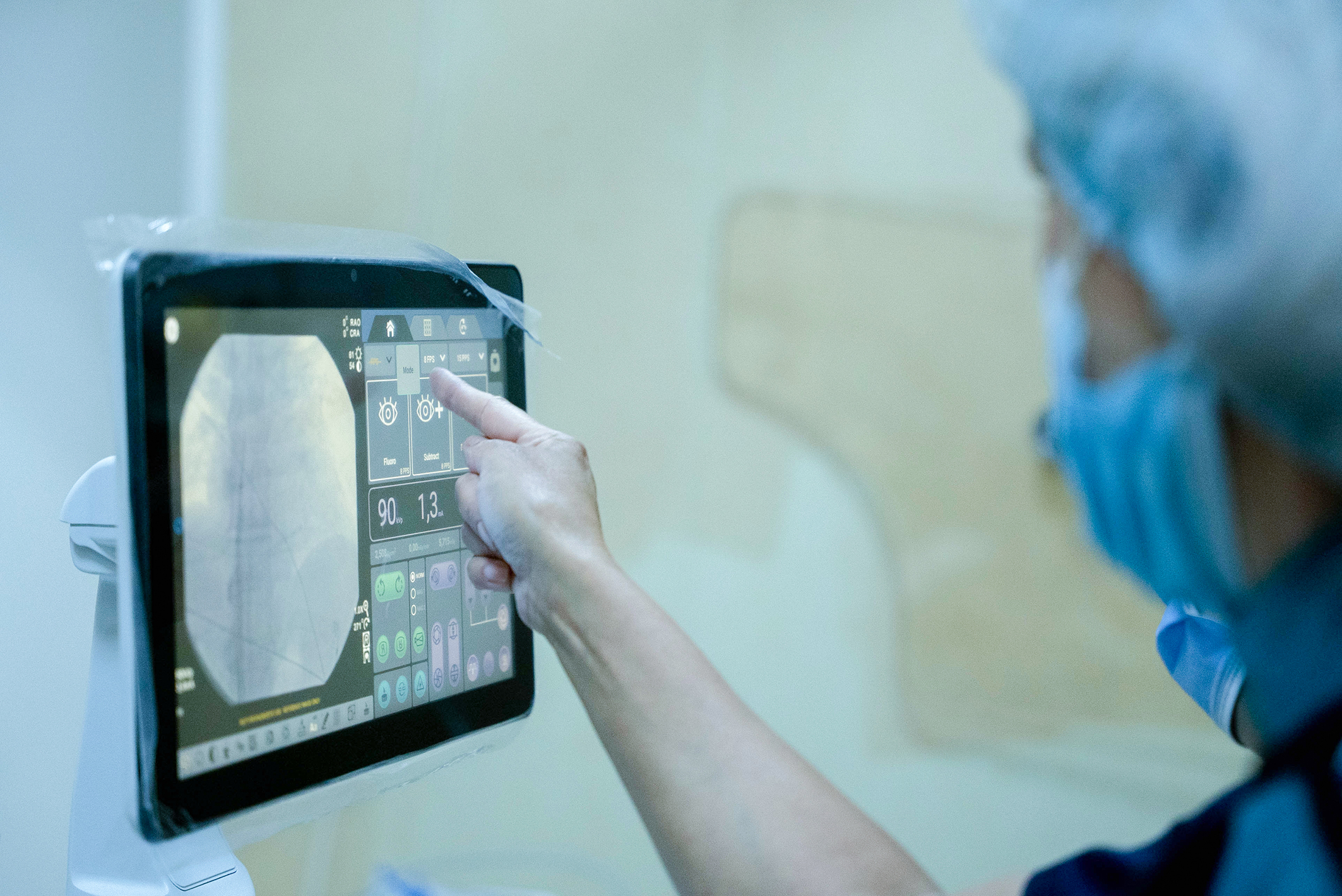
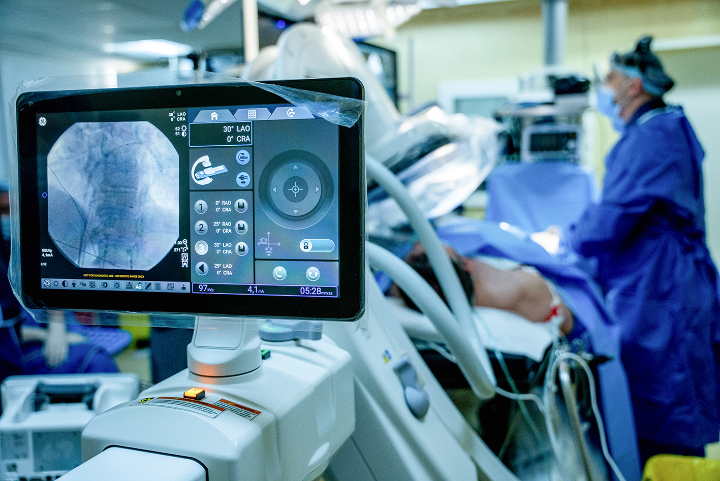
"The OEC Elite CFD significantly improves image quality while requiring lower doses of radiation compared to our previous C-arm. The decision to choose the OEC Elite CFD was also influenced by its maneuverability. The motorized model, in particular, makes it easier for both our team and the staff to rotate and control the C-arm. The OEC Touch control panel simplifies the process with just one button push to achieve projections such as Left Anterior Oblique (LAO) or Right Anterior Oblique (RAO)."
“OEC Elite CFD is very intuitive and very easy to use thanks to the motorization of the C-arm and the preset LAO and RAO positions.”
Dr. Sergiu-Nicolae Şipoş
You mentioned earlier that cryoablation is a complex procedure. Can you talk about the benefits that OEC Elite CFD brings to that procedure?
"The imaging needs for balloon cryoablation are somehow different from the conventional radio frequency ablation.
For conventional RF ablation, you need 2D visualization of the position of the catheters and also for the transseptal puncture, just to see where you are in the area of the cardiac silhouette.
The imaging protocol for balloon cryoablation differs somewhat from simple procedures. It demands better image quality because we have to perform a pulmonary vein venography. So, it’s not just about placing the catheters into the right cardiac chamber, but we also need to perform a good occlusion of the vein in order to treat it with a cryoballoon which comes deflated. At a certain point, we inflate this balloon, and we push it towards the ostium of the pulmonary veins to obtain total occlusion. To control it, we perform some contrast injection to check that we can’t see the pulmonary vein and its branches.
The success of the procedure relies on achieving a thorough occlusion.
Inadequate contact or contrast leakage around the balloon indicates poor occlusion, leading to a failed ablation. This is particularly critical in EP procedures, especially those targeting arrhythmogenic tissue around the ostium of the pulmonary vein. Such procedures demand high-quality images, precisely delivered by the OEC Elite CFD motorized Cardiac C-arm.
In our simple procedures, we always utilize low-dose fluoroscopy or a low frame rate. However, for complex procedures like cryoablation, we start with the low-dose mode and set the fluoroscopy at four pulses per second. If the image quality falls short, we incrementally adjust the dose by eliminating the low-dose mode and increasing the pulse rate. The motorized cardiac C-arm of the OEC Elite CFD simplifies this adjustment process.
It has been my instinctive approach to keep the radiation dose as low as possible. If the image quality is insufficient, we gradually escalate the dose, never commencing with medium or high doses. Most often, the image quality remains satisfactory with low-dose settings."
“The quality of the image is pretty good with low dose settings.”
Dr. Sergiu-Nicolae Şipoş
What other procedures are benefitting from your new OEC Elite CFD?
"There are other specific procedures we are doing with OEC Elite CFD that require very high-quality images. For instance, in the case of transvenous pacing, we employ specialized leads with small helices at their tips, which can be challenging to visualize. The OEC Elite CFD C-arm assists us in discerning the optimal positioning of the helices, crucial for ensuring a secure fixation between the lead and the myocardium.
The physiological pacing system approach, for instance, is a recent trend in pacing, that requires us to use delicate leads positioned in a very small area within the human heart. This precision is crucial to enable the lead to pace the normal conductive system accurately. Hence, our precision becomes paramount when working inside the patient’s heart.
Conventional cardiac resynchronization therapy (CRT) is a rather complex procedure which requires us to place a third lead outside the heart in the coronary sinus veins situated on the epicardial surface of the heart. To achieve this, it is essential to have a good venography and a good visibility on the tiny guidewire, which has a diameter of 0.014.
As described earlier, performing a transseptal puncture to cross the interatrial septum is another moment that demands a very good image."
“We need to have a good venography and a good visibility on the tiny guidewire that we use, the diameter of which is 0.014‘’. ”
Dr. Sergiu-Nicolae Şipoş
In your opinion what are the most advantageous features of the OEC Elite CFD motorized Cardiac C-arm?
“The high-quality images achieved at lower doses is obviously very important.”
Dr. Sergiu-Nicolae Şipoş
"The staff is aware that as they start the machine, they will select the ”low dose” mode just by pressing the ”low dose” button.
It is highly intuitive and user-friendly. The motorization of the OEC Elite CFD, along with preset positions, enhances its ease of use."
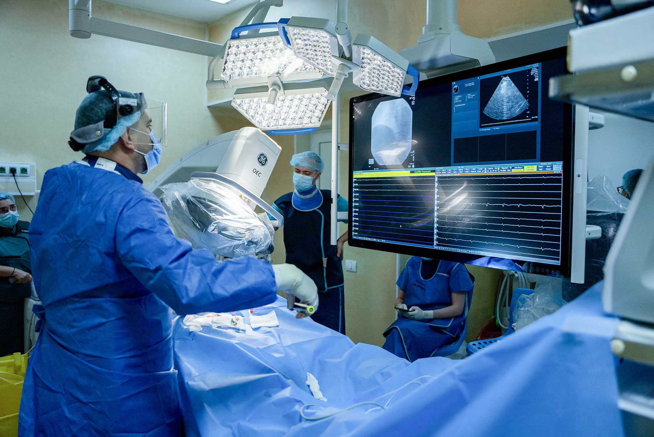
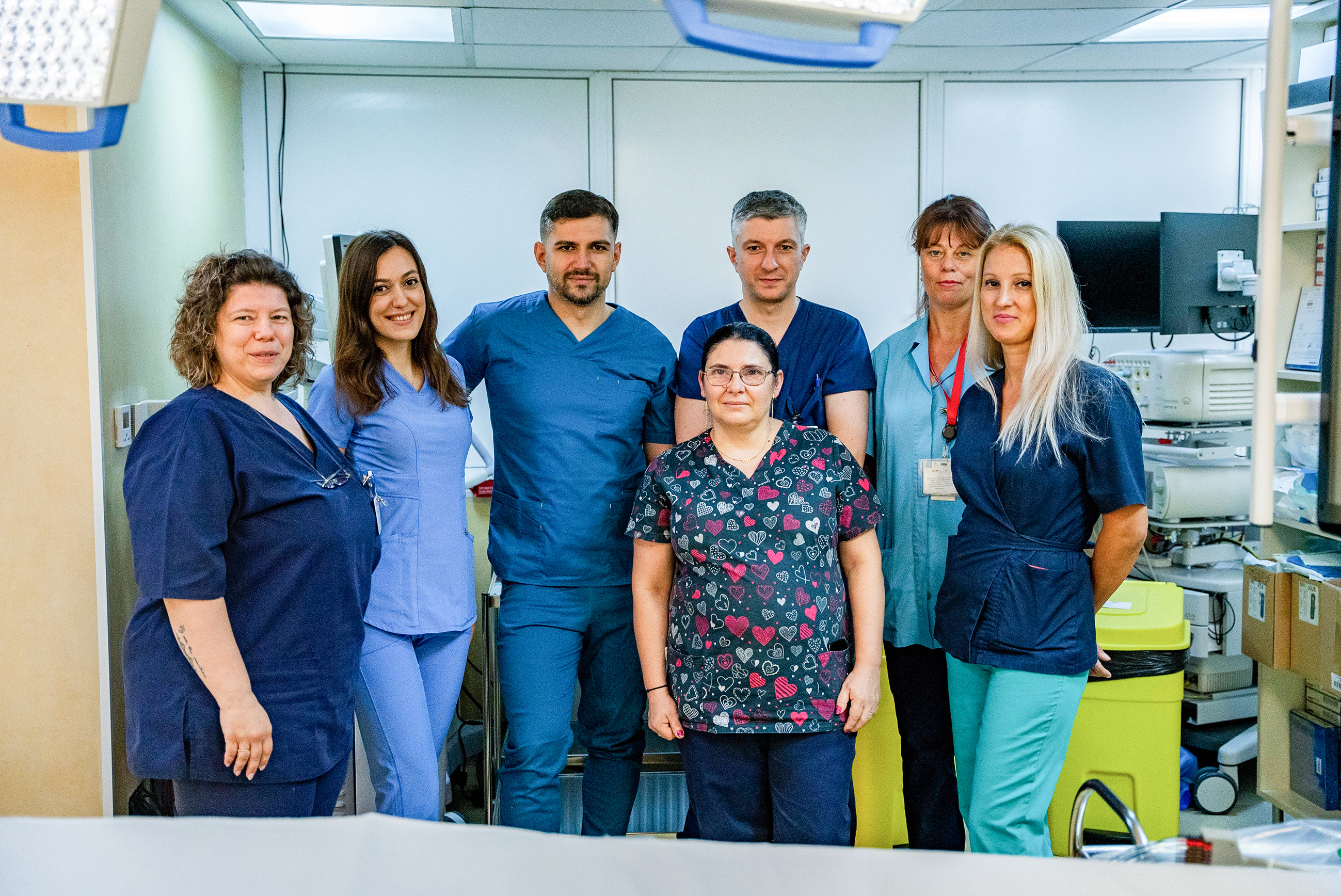
From left to right and back to front: Cristina Ungureanu (nurse), Claudia Nistor (resident Medical Doctor), Leonard Mandes (Medical Doctor), Sergiu Sipos (Medical Doctor), Laura Apreutesei (nurse), Violeta Mereuteanu (nurse), Florina Socol (nurse)
 |
Dr. Sergiu-Nicolae Şipoş is a Cardiologist and Interventional Rhythmologist at the EP and Pacing Department of Institute for Cardiovascular Diseases, hospital CC Iliescu, Bucharest, Romania. Dr. Şipoş has been a senior physician in cardiology since 2009 and an electrophysiologist for about ten years. He has a PhD in cardiology with a focus on cardiac arrhythmias and devices in heart failure. |
Balloon cryoablation of pulmonary veins (PV)
An electrophysiology (EP) procedure performed with OEC Elite CFD
Courtesy of Dr. Sergiu-Nicolae Şipoş, Cardiologist and Interventional Rhythmologist, EP and Pacing Department, Institute for Cardiovascular Diseases, hospital CC Iliescu, Bucharest, Romania.
Challenges
Balloon cryoablation of PV is an EP technique to achieve PV isolation (PVI) and restore a regular cardiac rhythm. The cryoablation balloon is positioned and pushed forward over the ostium of the PV to treat until complete occlusion is obtained. After that step, cryoablation is initiated resulting in the cauterization of the PV’s surroundings by freezing the cardiac cells.
One challenge of the procedure is to bring the cryoablation sheath from the right atrium (RA) to the left atrium (LA) of the heart via a transseptal puncture. Transseptal puncture might be a complex gesture that increases the need of fluoroscopic imaging.
Another challenge is to properly position the balloon over the vein. Confirmation is obtained through venograms requiring a good image quality in thick and moving anatomies.
The configuration and set up of OEC Elite CFD have been selected to address these challenges.
OEC Elite CFD configuration
The cathlab room is equipped with OEC Elite CFD in Cardiac configuration with the motorized drive C-arm and the 31 by 31 cm CMOS detector.
The motorized drive C-arm offers the possibility to preset and recall Left Anterior Oblique (LAO), Right Anterior Oblique (RAO) and cephalad-caudal angulations along the procedure from the Touch tablet (see Figure 1).
The deep Super C-arm of this configuration permits the installation of its mainframe at the extremity of the patient table to clear patient access while still allowing the detector to reach the cardiac area (see Figure 1).
The large size detector covers the anatomy of the heart which silhouette is used as a landmark by practitioners to guide their gestures during the procedure.
OEC Elite CFD set up
During the cryoablation procedure, the C-arm is positioned with 30-60° LAO angulation for the positioning of the balloon on the left PV (LPV), and 30-45° RAO angulation for the treatment of the right PV (RPV).
The imaging profile selected is Cardiac with the eNR correction to crisp the image of the tip of small catheters and guidewires (up to 0.014 inches of diameter) while in motion inside the beating cardiac chambers. To manage radiation exposure the fluoroscopy mode is set to low dose and pulse mode (4 pulses per second).
Conclusion
OEC Elite CFD provides efficient support with precise fluoroscopic imaging guidance while managing radiation dose exposure.
Figure 1: OEC Elite CFD C-arm is placed at the head of the patient. OEC Touch control panel shows the preset position menu.
Cryoablation of left pulmonary veins (LPV)
C-arm is angulated 45° LAO. Fluoroscopy mode is set to 4 pulses per second and low dose.
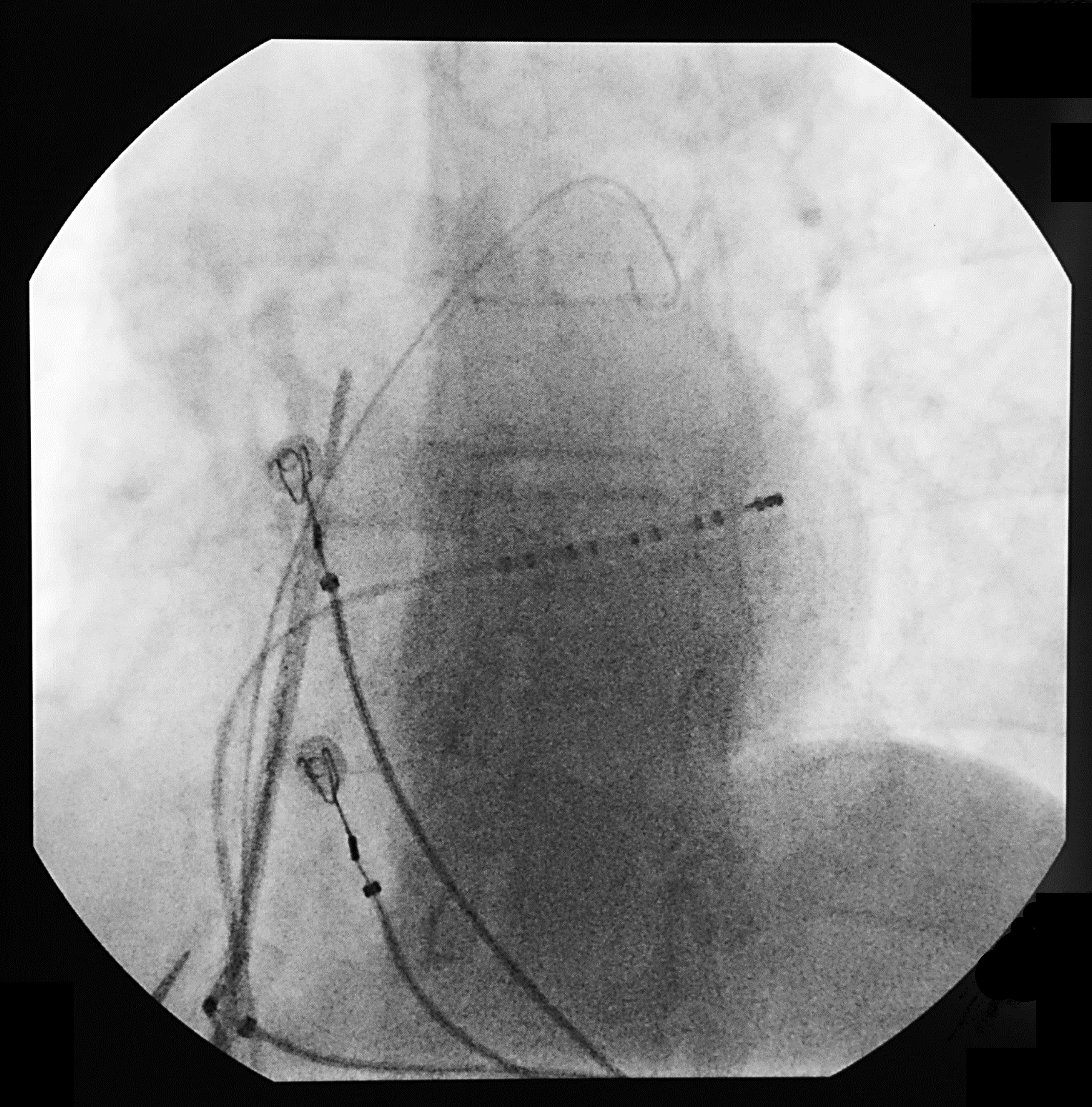
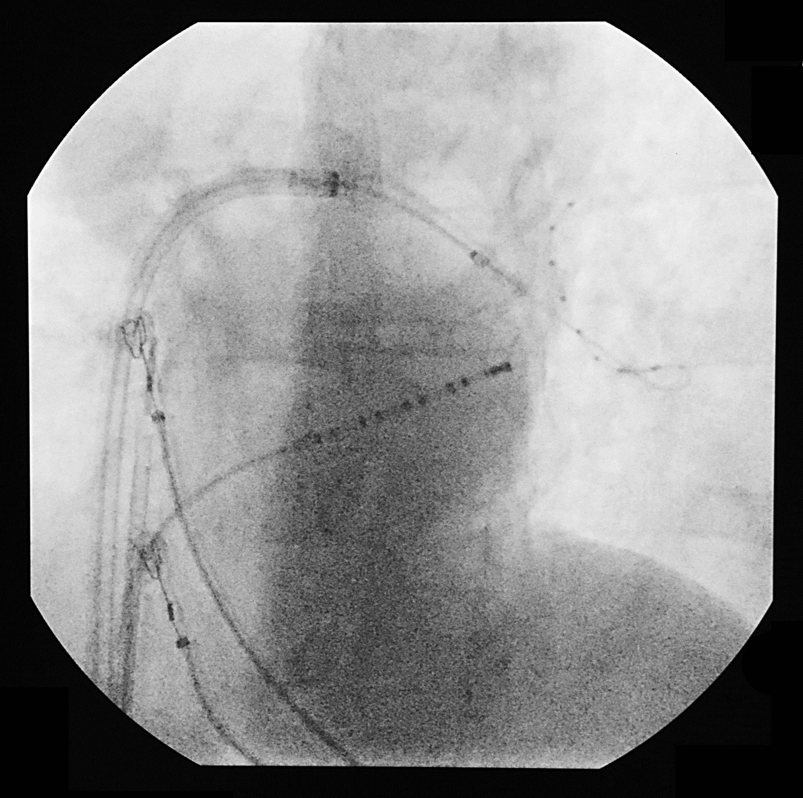
The guidewire is pushed from the RA to the LA through the fossa ovalis under fluoroscopic guidance. The full detector field of view is kept to observe the cardiac silhouette during the gesture (Figure 2). The transseptal dilatator and the sheath carrying the cryoablation balloon are advanced across the septum. The balloon is inflated and pushed forward to occlude the ostium of the PVs. Contrast media is injected to perform a venogram of the PV and its branches to control the proper occlusion of the ostium of the PV to treat by the balloon (Figure 3). The cryoablation balloon is moved inside the LA to modify its position (Figure 4) until the venogram confirms its proper positioning (Figure 5).
Figure 2
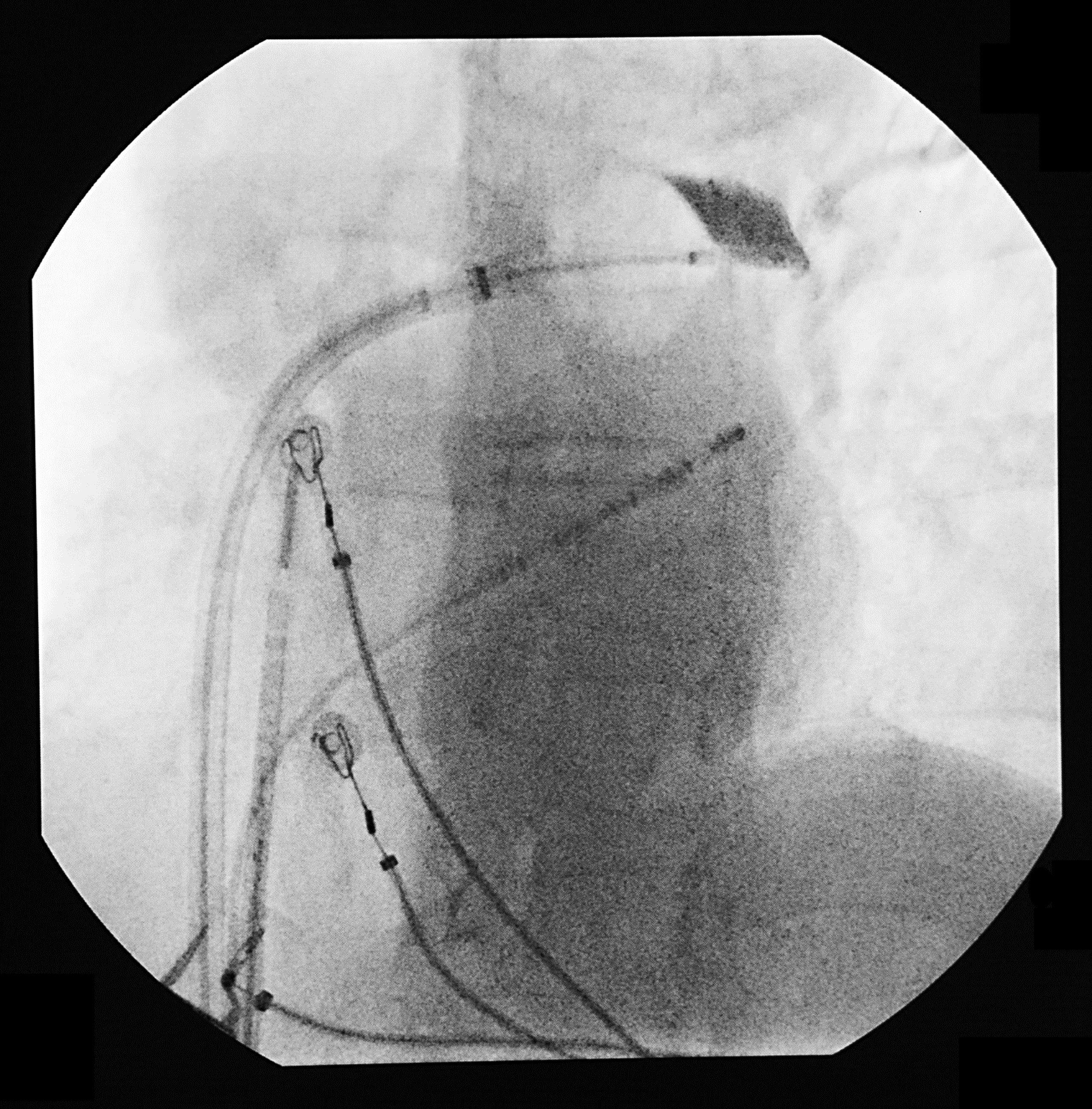
Figure 3
Figure 4
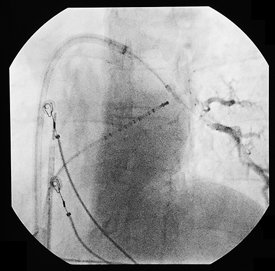
Figure 5
Cryoablation of right pulmonary veins (RPV)
C-arm is angulated 35° RAO. Same steps are repeated to insure the proper positioning of the balloon over the RPV (Figure 6).
Figure 6
The statements by GE’s customers described here are based on their own opinions and on results that were achieved in the customer’s unique setting. Since there is no “typical” hospital and many variables exist, i.e. hospital size, case mix, etc.. there can be no guarantee that other customers will achieve the same results. JB04054UK

