Experience with OEC 3D for endovascular aortic repair (EVAR)
With the courtesy of Pr. Jonathan Sobocinski, vascular surgeon, Head of Department of Vascular and Endovascular Surgery, Dr. Maxime Lebaz and Dr. Zoe Ribreau, vascular surgeons, Aortic Center, Lille University Hospital, France
EVAR: Infrarenal stent graft with left iliac branched device
Presented by Dr. Maxime Lebaz, Dr. Zoe Ribreau and Pr. Jonathan Sobocinski, vascular surgeons, Aortic Center, University Hospital Lille, France.
Clinical Aspect
Over the last decade, endovascular aortic repair has become the preferred surgical technique to treat aneurysm and dissection. Patients’ selection relies on strict anatomical criteria. The preoperative planning of the procedure includes careful analysis of the morphology of the aortic lesion and the ilio-femoral access. The technical feasibility and the stent graft sizing are then subsequently determined. The procedure is performed under fluoroscopy guidance, and the X-ray dose delivered throughout the procedure depends on the complexity of the navigation (calcification, tortuosity, diameter) and patients’ morphotype.
When such procedures are performed with a fixed imaging system, routine CBCT scans are completed at the end of the procedure to ensure proper integrity of the stent graft and to depict any conflict on the devices implanted. These intraprocedural acquisitions secure the quality of the arterial reconstruction and have proven reduction in early and late reinterventions1.
Technical Solution
OEC 3D is developed by GE HealthCare. OEC 3D is a mobile C-arm with 2D and 3D CBCT imaging using CMOS flat panel technology.
The OEC 3D C-arm provides a 19 cm x 19 cm x 19 cm high resolution 3D volume within 30 seconds of image acquisition, including up to 400 images with 3 different acquisition modes (Standard, High Definition and High Definition +). Integrated processing algorithms can smooth metal artifact (Metal Artifact Reduction) and reduce surrounding noise (enhanced Noise Reduction).
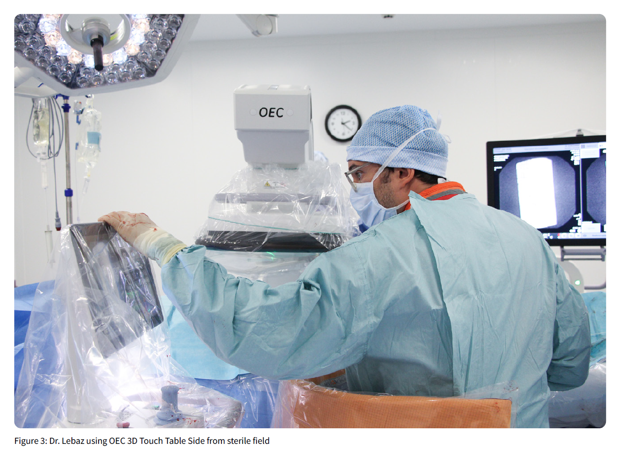
The user interface to plan and perform the 3D acquisition sweep (3D Setup Assistant) is easy to use: biplane lasers and a collision check step allow for a precise centering of the anatomy in the 3D volume. A 200° sweep of the C-arm collects 3D data in 30 seconds (3D data acquisition step). An additional 30 seconds of data reconstruction occur before display of volumetric images on the video monitor of the workstation.
The surgical table used is ImagiQ2TM (Stille AB, Solna Sweden).
The patient here is an 88 year-old with a large (80 mm) infrarenal abdominal aortic aneurysm extended to the left common iliac artery.
Case & Procedure
The treatment proposed was an endovascular exclusion of the aneurysm with an infrarenal stent graft in addition to a left iliac branched device to seal its left distal part within the external and the internal iliac arteries [Gore Excluder AAA Endoprosthesis, and Gore Excluder iliac branch endoprosthesis, W.L. Gore & Associates, Flagstaff, Arizona, USA].
The best projection to optimize the visualization of the origin of the lowest renal artery and iliac bifurcations were assessed from the preoperative computed tomography angiography (CTA).
The procedure was performed percutaneously under general anesthesia. The iliac branched device was first deployed associated with the bridging graft to ensure sealing in the internal iliac artery. Then, the positioning of the main bifurcated aortic body before deployment was
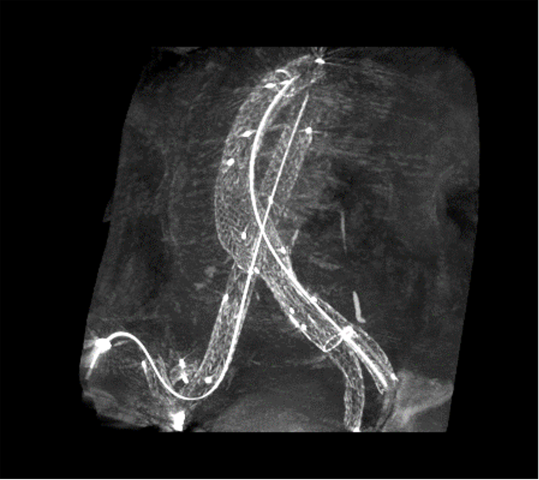
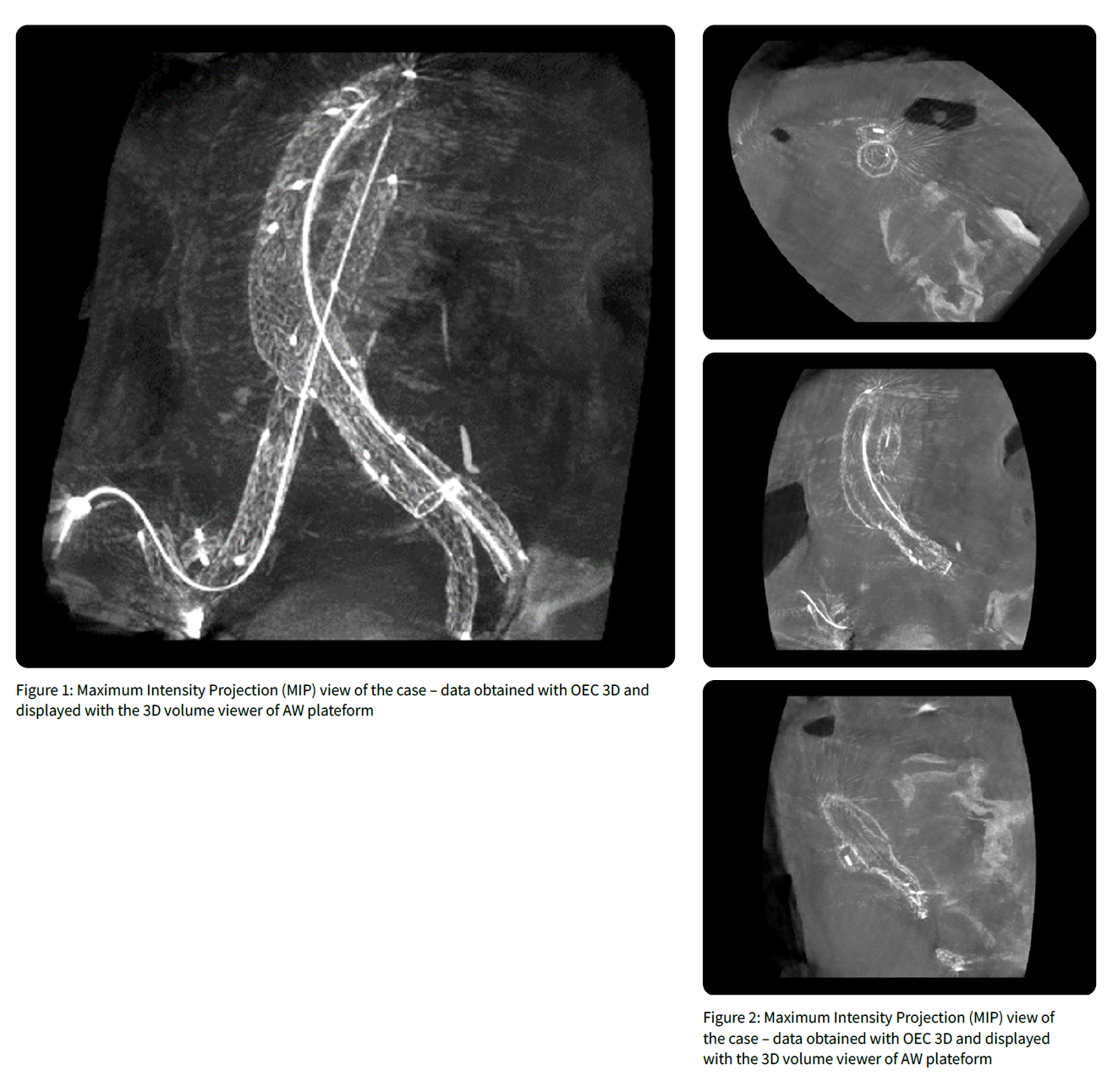
checked with a short angiogram of 7 cc (cubic centimeters) of contrast media at the origin of the lowest renal artery. The subsequent usual steps of the procedure were achieved without any particular issue and iliac limb extensions were deployed in order to ensure optimal overlap between graft components on the left handside and sufficient application within the native right common iliac artery. A final 2D DSA (digital subtracted angiography) with 25 cc of contrast media confirmed aneurysmal exclusion, the renal and hypogastric arteries and stent graft patency. A mobile CBCT scan with no injection was then completed. The entire procedure lasted 100 minutes. The total fluoroscopy time of the procedure was 19 minutes, with a total Dose Area Product (DAP) of 44.445 Gy.cm² and the total amount of contrast injected was 40 cc [Xenetix® 300mg/dl (Guerbet, France)].
Volumetric images acquired with OEC 3D at the end of the procedure
Specific featuring
The OEC 3D mobile CBCT C-arm is similar in terms of use and workflow to the OEC Elite
CFD C-arm. The entire 2D fluoroscopic imaging procedure was completed in low dose and pulsed mode (8 pulses per second (pps)). At the beginning of the procedure the automatic storage of all fluoroscopic acquisitions was activated (dynamic recording): it reduces the need of DSA acquisitions along the procedure.
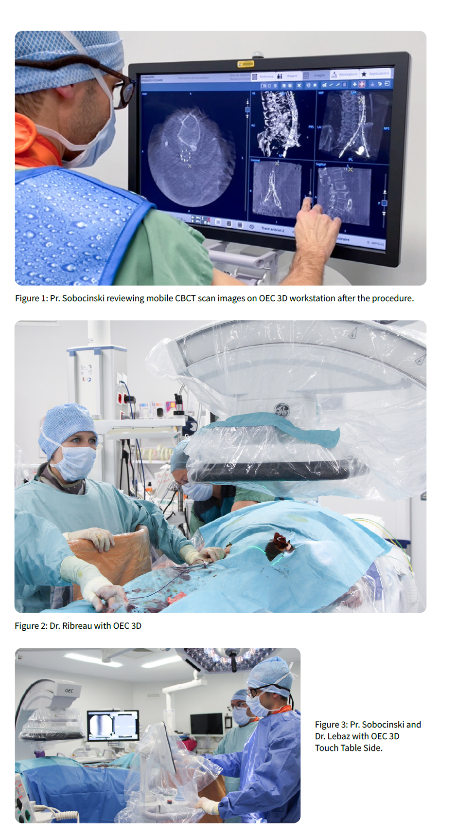
For the 3D acquisition, the ‘3D Setup Assistant’ software guides the user through 4 steps: patient orientation and imaging mode selection (step 1), centering the anatomy in the 3D volume (step 2), collision check (step 3), and 3D acquisition (step 4). After 30 seconds (duration of the 3D volume reconstruction) the volumetric images are displayed, and the Volume Viewer automatically opens with a suite of 3D imaging tools based on GE HealthCare AW image fabric.
The Volume Viewer displays 5 volumetric images on the monitor screens: cross sectional planes (axial, coronal, sagittal), volume rendering (VR) and maximum intensity projection (MIP).
The Volume Viewer’s tools enable the user to choose the display mode options and several tools for viewing images of cross-sectional planes (such as Multi Oblique Mode, scrolling through slices, slice thickness adjustment, window level, zoom, measurement, annotation and image storing). For VR and MIP images it also allows users to rotate the image.
Conclusion
Introducing the possibility of completing an intra operative mobile CBCT scan with OEC 3D offers additional safety during EVAR.
Moreover, the mobile CBCT scan feature in OEC 3D is an easy-to-use tool with short learning curve for surgeons.
The 3D images obtained provide a reliable assistance for checking the quality of the endovascular exclusion and might alleviate the postoperative imaging follow-up requirements.
Introducing CO2 in the aorto-iliac region
Courtesy of Dr. Maxime Lebaz, Dr. Zoe Ribreau and Pr. Jonathan Sobocinski, Vascular surgery, Aorta Center, University Hospital Lille, France.
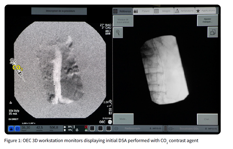
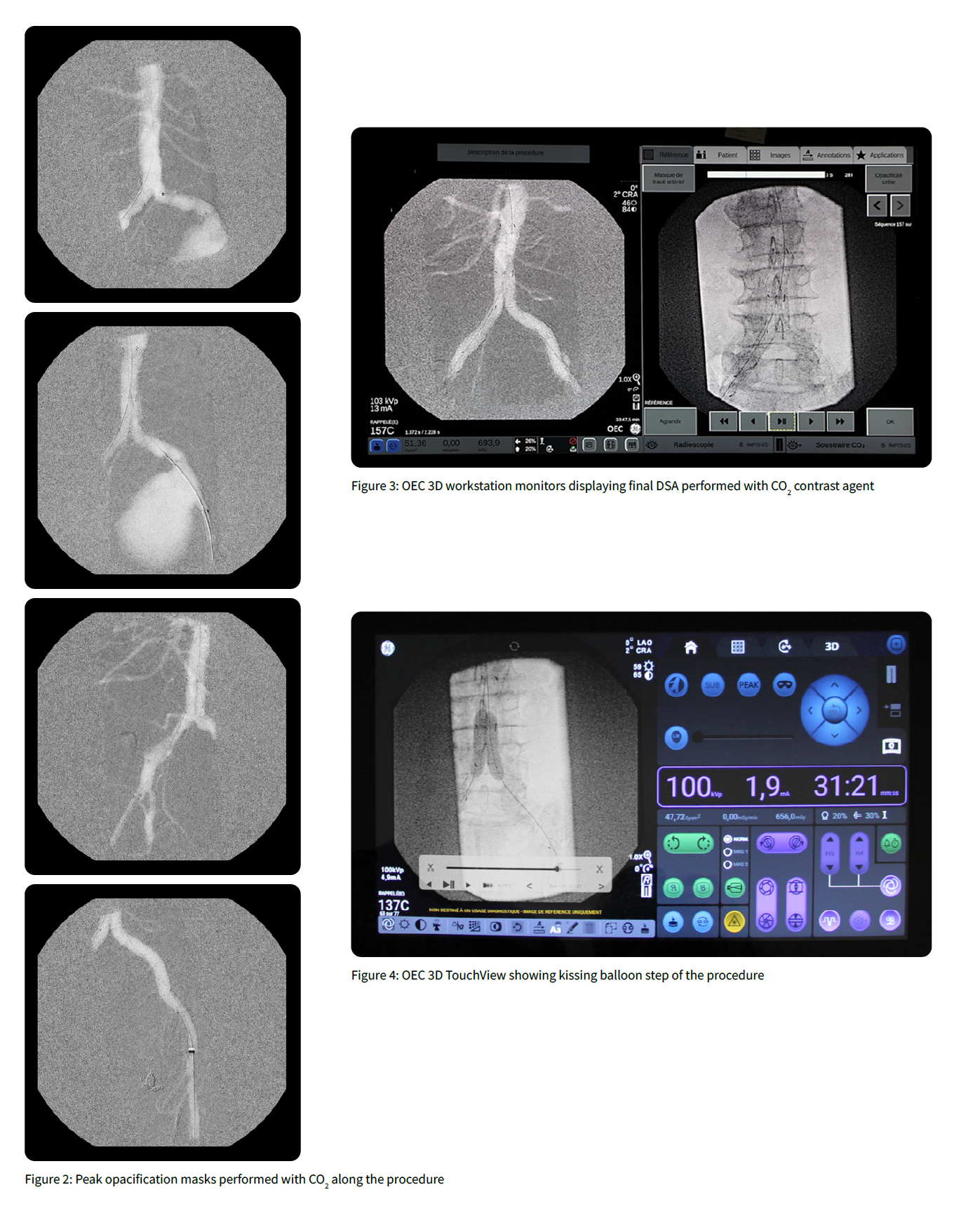
CO2 is a gas 20 times more soluble in blood than oxygen and can be used in endovascular procedures as a contrast agent to help in navigation and assess vessel patency. CO2 is eliminated by lungs and represent an alternative to iodized contrast agents in patients with renal dysfunction. CO2 injections can be associated with potential side effects reported as cerebral gas embolism and general discomfort. There is no specific dose threshold as long as an appropriate interval is maintained between two injections and the injection should be in the vessels below the diaphragm. The injection is performed using a dedicated automatic injector. Since CO2 is less dense than iodine contrast media (ICM), its X-ray absorption coefficient is lower, and requires a specific image processing in the different types of image acquisitions.
We present here the case of an octogenarian patient with known severe renal insufficiency with an estimated Glomerular Filtration Rate (eGFR) at 41 ml/ min where was incidentally diagnosed a large aneurysm (10 cm in diameter) in his left common and internal iliac arteries. Considering the location of the aneurysm and the patient’s medical condition, an endovascular exclusion guided with CO2 as a contrast agent was performed.
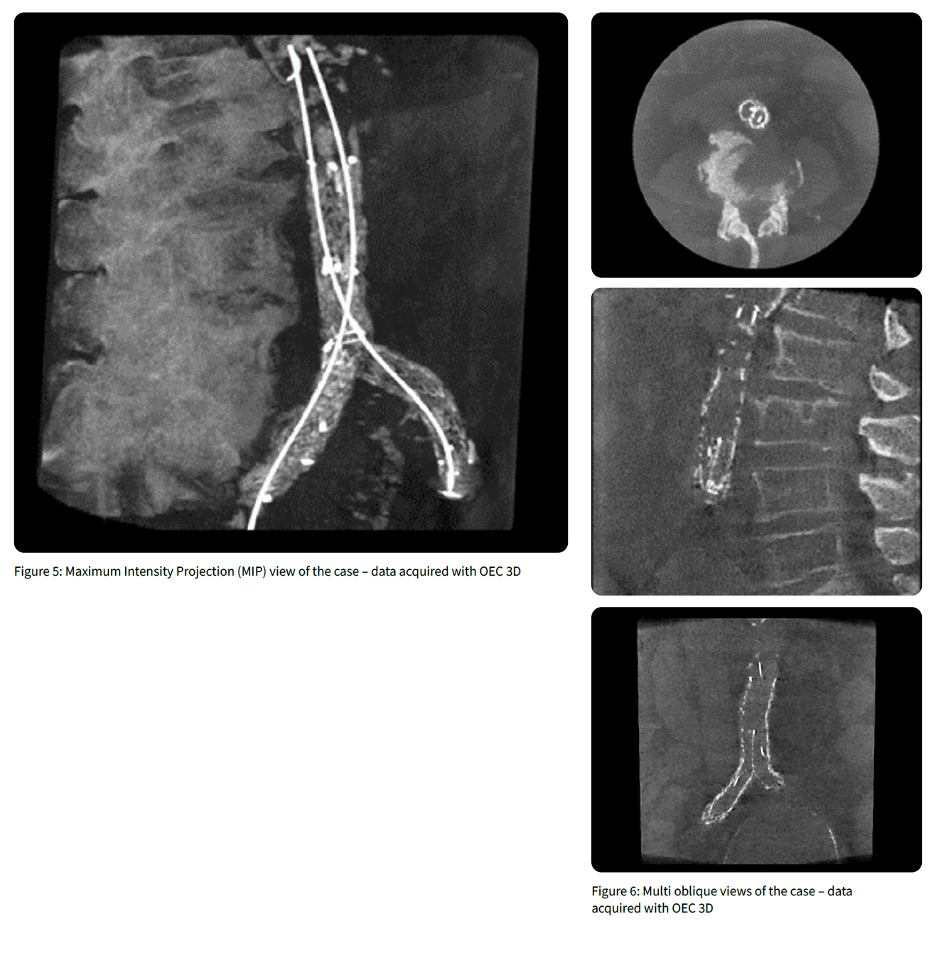
The procedure included the embolization of the distal branches of the left hypogastric artery and the implantation of a bifurcated infrarenal stent graft with a left external iliac extension covering the origin of the hypogastric artery. The embolization enclosed the implantation of an Amplatzer AVP2 plug (Abbott) in one of the main distal branches of the hypogastric and was completed by pushable coils in other small branches [Concerto coils (Medtronic)]. Subsequently, after visualization of the lowest renal artery with a CO2 aortography (volume and flowrate to be provided and administrated by the Angiodroid injector (Bologna, Italy)), a bifurcated infrarenal stent graft (RLT261412, Gore) was implanted. A final 2D angiography (25cc of ICM at a 15cc/sec) to assess the patency of the stent graft and potential perigraft leak was performed. A mobile CBCT scan using the high-definition mode of the OEC 3D was completed additionally and depicted constraints by the aorto iliac native bifurcation on both limbs of the stent graft requiring additional kissing stent (BeSmooth, Bentley).
The total procedure duration was 120 minutes, with 900 ml of CO2 injected and a cumulated dose area product of 27,25 Gy. cm²; the global amount of ICM injected was 25 cc.
The postoperative course was uneventful, and the eGFR at discharge was 48mL/min.
2D fluoroscopic imaging
Volumetric images acquired with OEC 3D at the end of the procedure
Pr. Sobocinski is a paid consultant for GE HealthCare and was compensated for participation in this testimonial. The statements by Pr. Sobocinski described here are based on his own opinions and on results that were achieved in his unique setting. Since there is no “typical” hospital and many variables exist, i.e. case mix, there can be no guarantee that other customers will achieve the same results.
OEC 3D is a mobile 3D/2D C-arm that provides intraprocedural mobile CBCT scan images and 2D fluoro images to enable visualization of anatomical structures during diagnostic, interventional, surgical, and minimally invasive surgery procedures such as endovascular procedures. JB28597XX

