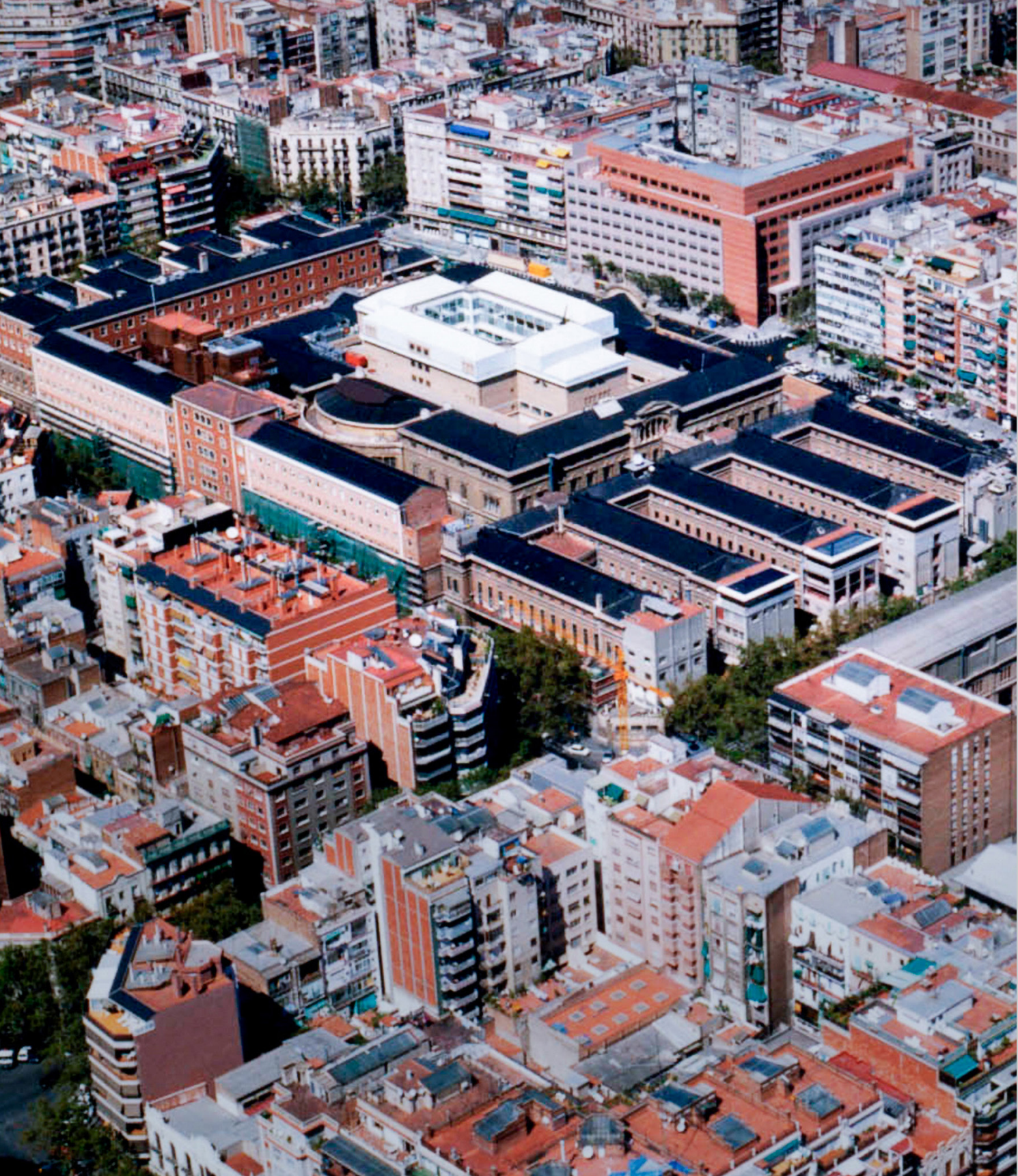
Hospital Clínic de Barcelona, Spain
The Hospital Clínic’s Cardiovascular Institute (ICCV) is devoted to treating cardiovascular diseases, with specialties that include vascular surgery, cardiovascular surgery and cardiology. Its interventional cardiology section has recently deployed GE HealthCare’s new 3DStent which is designed to address major stent imaging barriers.
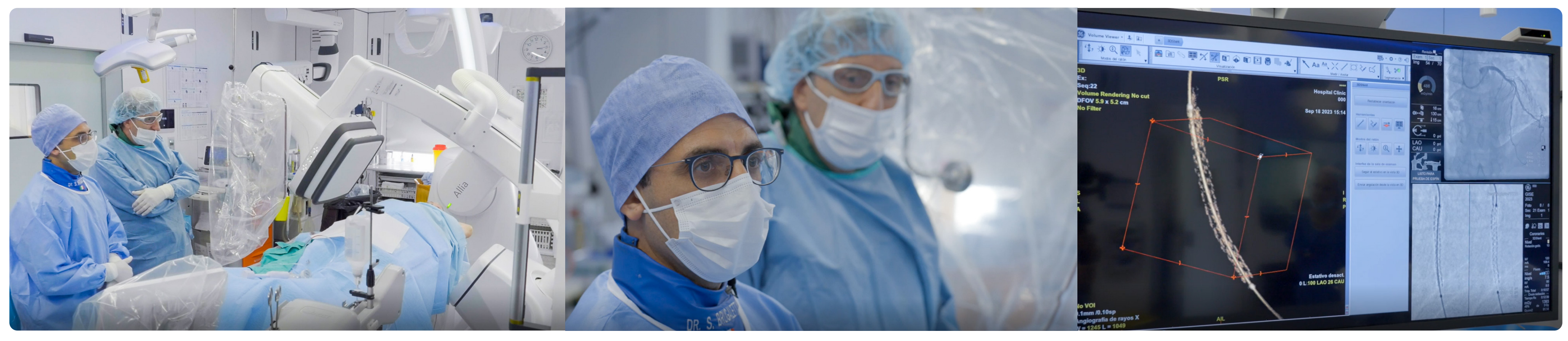
The Interventional Cardiology department at Hospital Clínic de Barcelona fully renovated their cathlabs and OR area in 2021. It is now equipped with two Innova IGS 520 with AutoRight and a hybrid room with the latest GE HealthCare platform, Allia IGS 730 with AutoRight. This hybrid operating theatre is mainly used to carry out structural heart procedures such as TAVI, LAAC, Mitral and Tricuspid valve interventions and percutaneous coronary interventions, among others.
In 2023, GE HealthCare launched 3DStent, an innovative tool which allows the intraprocedural visualization of a coronary stent in 3D and multi-slice images. The image is acquired through an automated rotation of the C-arm and thanks to the new CMCT (C-arm Motion compensated Computed Tomography) technology, 3DStent allows the clinician to see the stent from all angles without inserting an additional device or contrast.
It became CE marked in July 2023 and Hospital Clínic de Barcelona was the first site worldwide to use it in clinical practice.
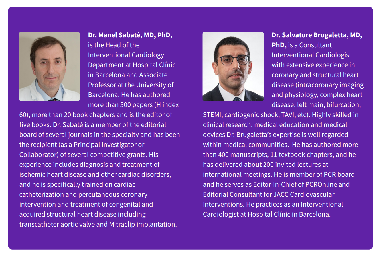
Dr Sabaté : In the coronary field we are facing more and more complex cases in our daily practice, what we call CHIP (Complex and High-Risk Interventional Procedures) cases, meaning that we have to deal with calcified plaques, bifurcations, coronary total occlusions, etc. In this scenario, it is critical to be able to assess the outcome of the procedure during the case. In these complex procedures, it is of the upmost importance that we’re using good imaging techniques and good equipment to ensure that we have a high-quality image of what we are doing.
As the first 3DStent users worldwide, Dr. Sabaté and Dr. Brugaletta tell us about their experience with this innovative intraprocedural 3D stent reconstruction technology in their clinical practice.
What is the impact of stent under expansion?
Dr Sabaté : Stent expansion is really important for the outcome of the patient. We cannot leave a stent underexpanded because it is a trigger for both restenosis and thrombosis. We have to make sure that the stents we have implanted are in the right position and well expanded inside the vessel wall, otherwise you will end up with problems for the patient down the road.
Dr Brugaletta : Stent expansion is one of the most important factors we consider during stent implantation. The expansion that we’re able to achieve when we implant a stent is directly related to the outcome of the procedure. We cannot leave a stent that’s not at least 80-90% expanded.
How are you dealing with stent expansion at Hospital Clínic?
Dr Sabaté : To make sure that the stent is well-apposed, well-expanded, we have several techniques. Of course, angiography is first, but with angiography alone we cannot be sure that the stent is optimally expanded. Another option we typically use is StentViz and we also use imaging techniques like intravascular ultrasound (IVUS) and optical coherence tomography (OCT). These are the typical techniques we use to assess expansion in our cathlab.
Dr Brugaletta : Usually when we do not see any calcium, we can see with StentViz or just with angiography if the stent is well expanded or not. But in cases where we have some calcification, tortuosity, or for example, a left main or a bifurcation, all these different angiographic features are strictly associated with bad outcomes. It’s especially important for these patients that we confirm a good stent expansion. We usually use intracoronary imaging, like IVUS or OCT, in order to ensure the correct expansion of the stent, not only in terms of percentage, but also in terms of square millimeter of area.
As the first 3DStent user worldwide, can you tell us what important information you can see during the procedure in the 3DStent images?
“We can see if there are areas of under expansion that may require post-dilation and we can also see calcified plaques behind the stent.”
Dr. Sabaté
Dr Sabaté : First, we’re looking at the expansion of the stent. That’s what we want to see with the 3D reconstruction. We can see the expansion of the entire stented area and, importantly, we can see if there are areas of under expansion that may require post-dilation with a non-compliant balloon, or other technique. We can also see calcified plaques behind the stent, which is important because these typically correspond to areas of under expansion. This is what we want to see with a 3D reconstruction of the stent using a non-invasive tool.
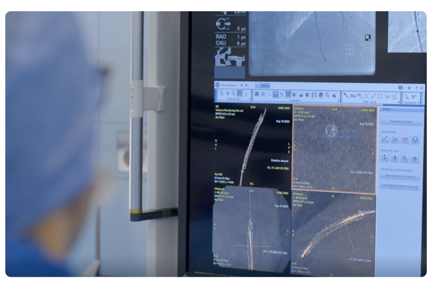
Dr Brugaletta : When I saw the images for the first time, I was quite surprised by the fact that we can see 3D also on the angio. We get lots of important information. The novelty is that we measure directly on the angiogram the minimum stent area of our stent, and this is something totally new. We usually use IVUS or OCT, but with 3DStent we save the cost of these catheters and assess what we need quite easily.
“We measure directly on the angiogram the minimum stent area of our stent, and this is totally new.” Dr. Brugaletta
The second bit of important information is the presence of calcification. Sometimes the vessel is calcified, and we can see where the calcifications surround the stent. We can understand if we are talking about a 360º calcification, or if it is just located in one quadrant. This information is very important because it tells us if there is plaque located behind the stent preventing it from being well expanded.
What do you like about 3DStent?
Dr Sabaté : You can measure the lumen of the stent, the length of the stent, the expansion of the stent, and you do not need to use an OCT catheter, so you can save some devices. We normally use OCT catheters, but not in every case and we can do 3DStent in every single case. I think this is the difference and may be the key factor of this new technology — you can use it in every single case in your daily practice.
Dr Brugaletta : What I like the most about 3DStent is actually how easy it is. All we need to do is to make sure that everything is set around the patient so we do not have any obstructions and that the x-ray can move freely around the patient. Then, it just takes 30 seconds to make the acquisition and 30 seconds of analysis. I find it very simple and intuitive to use.
How do you plan to integrate 3DStent in your clinical practice?
Dr Sabaté : What is important is to do a proper job for the patient and if you have a tool which only within 1-2 minutes can assess the entire expansion of the stent and the result of the PCI, I think you have to use it. When you are used to it, you will use it in every single case. I do not see any exceptions for not using it.
“It can easily be done in 100% of PCI.” Dr. Brugaletta
Dr Brugaletta : In all your procedures 3DStent makes it very easy to check stent expansion. Every time that you have a complex procedure, and want to guide that procedure by angio, you can do the 3D reconstruction. It may also save the cost of an imaging catheter if you don’t want to open it or if you have some OCT fatigue. Especially after the procedure, if you have used the catheter several times, you can use the 3D reconstruction just to check that the stent is well expanded and that you have done a good job instead of opening a new OCT catheter.
How do you think 3DStent will impact the interventional cardiology segment?
Dr Sabaté : We are an early user of this technology, but if we can compare it with invasive or catheter-based technologies like IVUS or OCT and it is comparable, we may save some catheters. So, at some point it may replace some catheters, may save some money we can use it in almost every single case.
Dr Brugaletta : In my opinion, it will have a positive impact. Of course, when we have something new in the lab, in our profession, we have to learn all of the utilities and software but once we do, it’s important that the new technology is easier to use compared to what it’s replacing and also less expensive than what we already have. 3DStent addresses both because it is quite easy to use and it’s also less expensive. This will impact our practice because it can easily be done in 100% of PCI.
Can you tell us about your partnership with GE HealthCare?
Dr Sabaté : I am really happy to be part of the launch of this new technology coming from GE HealthCare, to partner with them, and help improve this technology. I am pretty sure this is just a version 1.0 and in the future we will have new versions that will make our lives and the lives of our patients easier.
3DStent solution includes Allia™ system, 3DXR and Volume Viewer Innova and requires AW workstation with Volume Viewer. These applications are sold separately. Available on Allia™ IGS 5 with 20cm or 30cm detector and Allia™ IGS 7 with 30cm detector. Commercial availability of GE HealthCare medical systems is subject to meeting local requirements in a given country or region. Contact a GE HealthCare Representative for more information. Intended for healthcare professionals only. The statements described here are based on their own opinion and on results that were achieved in their unique setting. Results may vary. JB07563XE